Rewriting the Rules for Brain Bleeds: What INTERACT3 Taught Us

Spontaneous intracerebral haemorrhage has long carried an air of clinical resignation, the diagnosis that quietly lowered everyone’s expectations before treatment had even begun. INTERACT3, published in The Lancet in May 2023, unsettled that fatalism. It changed not a drug but a way of thinking, and it has since become impossible to discuss acute haemorrhagic stroke without reference to it.

The Trial in Brief

INTERACT3 was a pragmatic, international stepped-wedge cluster-randomised trial enrolling 7,036 patients across 121 hospitals, most of them in low- and middle-income countries. Instead of testing a single molecule, it asked whether a coordinated care bundle, begun within hours of the bleed, could alter the course of the illness. The bundle drew together four physiologically targeted actions: intensive systolic blood pressure lowering to a target below 140 mmHg; strict glucose control (6.1–7.8 mmol/L in patients without diabetes, 7.8–10.0 mmol/L in those with it); fever control holding temperature at or below 37.5°C; and rapid reversal of anticoagulation to an INR below 1.5.

What the Numbers Showed

The primary outcome was functional recovery, measured by a modified Rankin Scale shift analysis at six months. The bundle came out ahead, with a common odds ratio of 0.86 (95% CI 0.76–0.97; p=0.015). The advantage extended beyond function. Mortality fell from 17.0% to 14.1%, and serious adverse events dropped from 20.1% to 16.0%. In a disease so often treated with a shrug, these are shifts of real consequence.

The Story of Secondary Injury

The physiology explains why. Outcome after an intracerebral haemorrhage is shaped less by the original bleed than by the secondary injury that follows, and each element of the bundle disarms a different, time-sensitive part of that cascade.

The first threat is early haematoma expansion, which occurs in roughly a third of patients within the opening hours and responds to blood pressure. That is the rationale for rapid, controlled lowering of systolic pressure. The trial’s smooth, protocol-guided titration also mattered: it likely spared patients the falls in renal and cerebral perfusion that may have blunted the effect in earlier blood-pressure trials such as ATACH-2.

Alongside pressure sit hyperglycaemia and fever, each an independent driver of perihaematomal oedema, oxidative stress, and a faltering penumbral metabolism. Anticoagulation-associated bleeds carry the highest expansion risk of all, which places rapid reversal among the most valuable components of the bundle.

The deeper lesson runs beneath the individual parts. None of these interventions had previously shown a convincing functional benefit on its own. Delivered together, early and in concert, they moved patients measurably towards independence. The whole outperformed the sum of its parts.

From Nihilism to a Protocol Worth Running

This reframes the disease. Intracerebral haemorrhage moves from a condition met with therapeutic nihilism to an actionable, protocol-driven emergency, one that sits naturally beside the bundled approaches already familiar from sepsis and STEMI care. For the neuroanaesthesiologist and neurointensivist, the implication is concrete: systems-level implementation — standing order sets, rapid-reversal pathways, and pre-specified physiological targets switched on in the emergency department and carried through into the operating room — achieves what isolated pharmacological effort has not.

Reading the Evidence Honestly

Two caveats temper the enthusiasm. The stepped-wedge design is vulnerable to temporal confounding and cannot, by its structure, isolate which single element carried the benefit, which means the bundle should be adopted as a complete package rather than dismantled into favoured parts. And because most patients were recruited in lower-resource settings, questions of external validity and generalisability deserve open acknowledgement rather than quiet assumption.

These limits notwithstanding, INTERACT3 is well positioned to shape forthcoming AHA/ASA and European Stroke Organisation guidance. It establishes early, multimodal physiological control as the emerging standard of care in acute intracerebral haemorrhage.


Sources: INTERACT3, The Lancet (2023) · INTERACT3 full text, PMC
Reading the Pupil Anew: Automated Pupillometry in Neurological Monitoring

For over a century, the pupillary examination has rested on a simple ritual: a clinician, a penlight, and a judgement rendered in an instant — is the pupil brisk, sluggish, or unresponsive? It is among the most venerable of bedside assessments, and also among the least dependable. When clinicians examine the same patient, their agreement on whether the pupils are unequal falls below fifty percent, and the unaided eye is wholly incapable of detecting the subtle, incremental changes that so often precede deterioration.

Automated pupillometry offers a more rigorous alternative. A calibrated flash of light is delivered, the pupil’s response recorded in fine detail, and the result distilled into a single value — the Neurological Pupil index (NPi), scaled from 0 to 5, with anything below 3 flagged as abnormal. In shining light and measuring the constriction that follows, the device tests the full pupillary light reflex — the afferent limb carried by the optic nerve (cranial nerve II) that senses the light, and the efferent limb carried by the parasympathetic fibres of the oculomotor nerve (cranial nerve III) that drives constriction. Its warning power rests chiefly on the efferent (CN III) pathway, which is exquisitely sensitive to rising intracranial pressure and is compressed early as the brain begins to herniate. A gradually falling NPi can therefore signal catastrophe hours before the dreaded “blown pupil” appears. Measured every four to six hours, it now helps anticipate dangerous swelling after stroke, head injury, and surgery, and — via the 2025 NPi-Connect system — feeds straight into the medical record as a trackable vital sign independent of who holds the light.

The supporting evidence continues to accumulate. Automated pupillometry is now employed to anticipate dangerous brain swelling in stroke, with measurements taken every four to six hours to follow the trajectory rather than a single isolated reading. The same principle has proven valuable following traumatic brain injury and neurosurgery, where a falling NPi identifies patients on the verge of decline. In early 2025, a system known as NPi-Connect began transmitting these readings directly into the electronic medical record, establishing the NPi as a properly documented, trackable vital sign that no longer depends upon the individual performing the examination.

The limitations, however, deserve candid acknowledgement. Commonly used sedatives and analgesics — opioids and dexmedetomidine among them — blunt the pupillary response, and recent work demonstrates that the depth of sedation can meaningfully distort the reading. Iris pigmentation, prior ocular surgery, direct orbital trauma, and certain neuromuscular conditions may all degrade its accuracy, and the measurement interrogates only a single nerve pathway, offering no insight into the wider brain. The four-to-six-hour intervals between readings likewise leave unmonitored windows.

The most compelling horizon lies in estimating the pressure within the skull without breaching it at all. Emerging systems infer that pressure from the faint pulsatile expansion of the skull, from gentle microwave or near-infrared signals, or from ultrasound of the sheath surrounding the optic nerve. One such approach now falls within roughly 3 mmHg of the true value, achieving accuracy to within a few points in approximately 72 percent of cases. Combining these signals — the pupillary index, a non-invasive pressure estimate, and the optic nerve measurement — offers a credible route toward sparing many patients an invasive monitor placed through the skull. For the present, the invasive monitor retains its primacy, and the prudent posture is to regard pupillometry as a valuable early-warning companion rather than the sole arbiter of a swelling brain.

Bedside Weaning Assessment for Myasthenia Gravis

Bedside Test Procedure Normal / Reassuring Value Clinical Significance (Red Flag)
Clinical Assessment Observe for signs of respiratory distress and bulbar weakness (speech, swallowing). No accessory muscle use, effortless breathing, strong cough, clear voice. Tachypnea, accessory muscle use, paradoxical breathing, weak “boggy” cough, slurred/fading voice.
Vital Capacity (VC) Patient takes a maximal inhalation and exhales fully into a bedside spirometer. > 20 mL/kg (ideal body weight) < 15-20 mL/kg, or a declining trend during the breathing trial.
Negative Inspiratory Force (NIF) / MIP Patient makes a maximal inspiratory effort against an occluded airway for ~20 seconds. More negative than -30 cm H₂O Less negative than -30 cm H₂O (e.g., -25, -20), or a worsening trend.
Single Breath Count Patient takes a deep breath and counts aloud steadily (e.g., “1-one-thousand, 2-one-thousand…”). > 25 < 15-20. Indicates severely reduced vital capacity.
Head Lift Test Patient lies supine and lifts their head off the bed, holding the position as long as possible. > 30 seconds < 20 seconds. Correlates with significant diaphragmatic weakness.
Breath-Holding Time After a maximal inhalation, the patient holds their breath for as long as possible. > 30 seconds < 20 seconds. Suggests poor respiratory reserve.

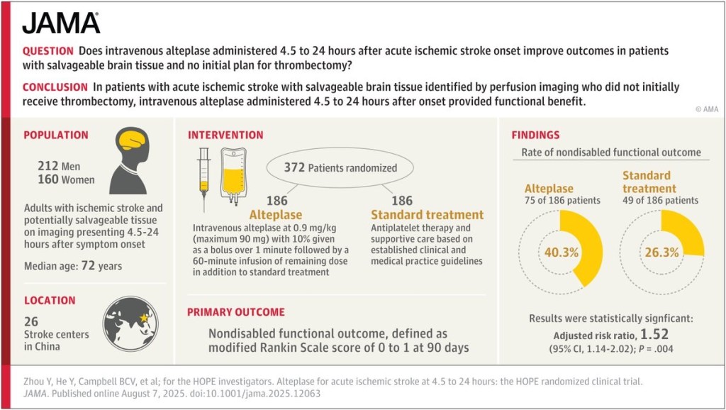
Does intravenous alteplase administered 4.5 to 24 hours after acute ischemic stroke onset improve outcomes?