How Much Blood Does an Injured Brain Need? What do HEMOTION and TRAIN tell?

The messages

For years, ICUs treated anemia in brain-injured patients like anemia in anyone else: transfuse only when hemoglobin falls to about 70 g/L. Two large 2024 randomized trials HEMOTION (NEJM) and TRAIN (JAMA) have overturned that habit, marking the biggest shift in neurocritical-care transfusion practice in a decade.

Why the injured brain is different

Injured brain tissue has almost no reserve to pull extra oxygen from blood or raise blood flow in the vulnerable “penumbra” around the injury. So anemia isn’t just a lab number here it becomes a direct driver of secondary ischemic injury at hemoglobin levels the rest of the body tolerates. Both trials tested whether keeping hemoglobin higher protects the brain.

HEMOTION: a strong signal, just short of significance

Adults with moderate-to-severe traumatic brain injury and anemia were randomized to a liberal strategy (transfuse at Hb <100 g/L) or restrictive (<70 g/L), with unfavorable Glasgow Outcome Scale “Extended (GOS-E) at six months as the primary outcome. Unfavorable outcomes: 68.4% liberal vs 73.5% restrictive  a ~5-point improvement favoring liberal, but not statistically significant. A “negative” trial that still leans one way.

TRAIN: larger, broader, clearly positive

Bigger and wider  850 patients, 72 ICUs, 22 countries, including traumatic brain injury plus subarachnoid and intracerebral hemorrhage. Liberal trigger <9 g/dL vs restrictive <7 g/dL, GOS-E at 180 days. Liberal was clearly better: 62.6% vs 72.6% unfavorable, an absolute risk difference of 10.0%, with no excess clotting or lung injury.

Do they really disagree? No.

The trials are concordant in direction, differing only in power and breadth. HEMOTION’s confidence interval includes TRAIN’s result, and both favor transfusing sooner. The likely mechanism is better cerebral oxygen delivery at the microvascular level supported by benefit appearing in functional neurologic recovery rather than survival.

What this means at the bedside

Shift toward a liberal threshold of ~90-100 g/L, individualized by evidence of brain-tissue compromise (brain-tissue oxygen tension, an evolving infarct, vasospasm risk) rather than a rigid number. A 70 g/L default is now hard to defend in structural brain injury. Two caveats: neither trial showed a mortality benefit (the case rests on function), and both were underpowered for disease subgroups. The open frontier is titrating transfusion to real-time cerebral oximetry or brain-tissue oxygen monitoring rather than a single systemic haemoglobin value.
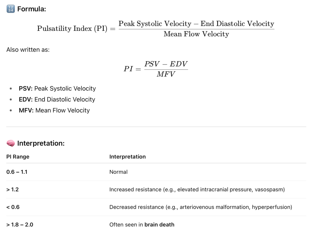
Pulsatility Index (PI)

The TCD Pulsatility Index (PI) is a parameter used in Transcranial Doppler (TCD) ultrasonography to evaluate the resistance to blood flow in cerebral vessels. It’s commonly used to assess cerebral hemodynamics, especially in patients with conditions like stroke, traumatic brain injury, hydrocephalus, and brain death.

📌 Clinical Uses of PI in TCD:

  • Elevated ICP (Intracranial Pressure): Higher PI suggests rising ICP.
  • Vasospasm detection in subarachnoid hemorrhage.
  • Brain death evaluation: Very high PI or absent diastolic flow.
  • Monitoring cerebral autoregulation.
  • Hydrocephalus assessment.

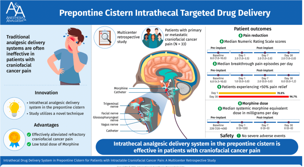
Intrathecal Drug Delivery System in Prepontine Cistern for Craniofacial Cancer Pain

Placing the catheter tip of an intrathecal morphine pump into the prepontine cistern could effectively relieve refractory craniofacial cancer pain with an extremely low total morphine dose requirement and few adverse events. This procedure could be considered in patients with severe refractory craniofacial cancer pain. (Anesth Analg 2025;141:255–63)

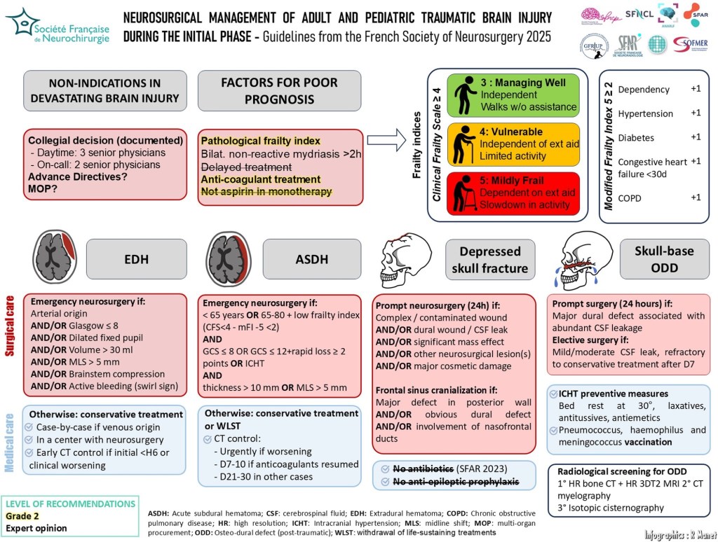
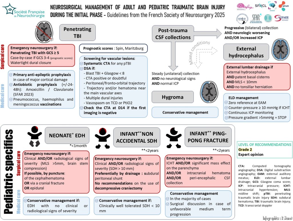
Neurosurgical management of the acute phase of adult and pediatric traumatic brain injury: 2025 guidelines of the French Society of Neurosurgery

Blood Transfusion in Pediatric Neurosurgeries: A Practical Guide


Introduction

Blood transfusion in pediatric neurosurgery, particularly for procedures like craniosynostosis repair, requires a careful and calculated approach. The unique physiological characteristics of children—especially infants—demand precise blood volume estimationloss assessment, and transfusion decision-making to ensure safety and optimal outcomes.

Below are key calculations and considerations when planning for blood transfusion in pediatric neurosurgical settings:


1. Estimated Blood Volume (EBV)

Understanding the Estimated Blood Volume (EBV) is essential for predicting transfusion needs.

Age GroupEstimated Blood Volume (ml/kg)
Preterm neonates90–100
Full-term neonates85–90
Infants (<12 months)80
Children (>12 months)75

For example, in craniosynostosis surgeries, which often involve infants, use 80 ml/kg; for children like a 5-year-old patient, use 75 ml/kg.


2. Maximum Allowable Blood Loss (MABL)

To guide intraoperative transfusion decisions, MABL is calculated as:

MABL = EBV × (Starting Hct – Target Hct) / Starting Hct

This helps determine the volume of blood a child can safely lose before transfusion becomes necessary.


3. Packed Red Blood Cell (PRBC) Transfusion Volume

To avoid over-transfusion:

PRBC Volume (mL) = Weight (kg) × Desired Hb rise (gm/dL) × Transfusion Factor

Where:

  • Transfusion Factor = 3 / PRBC Hct
    (Typical PRBC Hct = 0.60–0.65)

Example: 10 mL/kg of PRBC with Hct 60% gives ~2 gm/dL rise in Hb.


4. Methods of Estimating Blood Loss

Estimating intraoperative blood loss in neurosurgery can be difficult due to hidden bleeding. Use a combination of the following:

  • Visual assessment of the field
  • Calibrated suction canister readings
  • Weighing surgical sponges
  • Serial haematocrit levels from ABG
  • Thromboelastography (TEG) or ROTEM for coagulation monitoring

Note: Blood may be concealed under drapes or on instruments—constant vigilance is critical.


5. Factors Influencing Transfusion Decisions

There is no universal transfusion trigger; decisions must be individualized based on:

  • Preoperative haematocrit and baseline haemoglobin
  • Child’s weight and age
  • Surgical pathology (e.g., craniosynostosis often involves blood loss >20–500% of EBV)
  • Comorbidities affecting oxygen delivery (e.g., cyanotic heart disease)

Transfusion Thresholds and Recommendations

ScenarioTransfusion Recommendation
Hb ≥ 7 gm/dL and stableNo transfusion needed
Hb < 5 gm/dL or critically illTransfusion indicated
Hb 5–7 gm/dLIndividualized decision
Target Hb after transfusion7.0 – 9.5 gm/dL
Minimum Hct for craniotomy25%
Optimum cerebral oxygen deliveryHct ~30%
Acute brain injury (e.g., trauma)Transfuse if Hb 7–10 gm/dL
Massive transfusion (>50% EBV in 3 hrs or 100% in 24 hrs)Use PRBC : FFP : Platelet = 2 : 1 : 1

Summary

In pediatric neurosurgery, particularly in high-risk procedures like craniosynostosis repair, blood transfusion must be:

✅ Carefully calculated using weight-based formulas
✅ Guided by clinical condition, not just haemoglobin numbers
✅ Continuously reassessed using haematocrit, ABG, and coagulation studies
✅ Supported by a multidisciplinary team for timely intervention

By integrating these evidence-based parameters into your intraoperative workflow, you can significantly improve transfusion safety and patient outcomes.


Stay updated. Stay meticulous. Pediatric neurosurgery demands nothing less.