LUMBAR PLEXUS BLOCK

Anatomy of lumbar plexus?

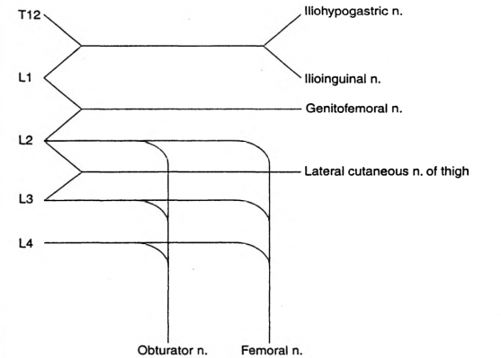
The lumbar plexus is formed by the anterior primary rami of L1 to L4 (Sometimes T12 also may contribute)
L1 forms the iliohypogastric and ilioinguinal nerves
It also gives a contribution to the formation of the genitofemoral nerve
L2 forms the lateral cutaneous nerve of thigh, along with L3
The obturator and femoral nerves are formed by contributions from L2,L3,L4

What are the indications?

Surgeries involving hip/thigh/upper leg/trauma
In conjunction with sciatic nerve or sacral plexus block
Cancer pain arising from hip or upper femur
Sympathetic block also helps in ischemic pain and CRPS

Which approach will you use?

I will use the 3 in 1 approach that aims to block the femoral, obturator and lateral cutaneous nerves with a low anterior approach. The patient is in the supine position.I will use a nerve stimulator. Using a 50 mm insulated needle, I will puncture the skin at a point 1 cm lateral to the femoral pulse and 2 cm below the inguinal ligament, at 45 degrees to skin and directed proximally and parallel to the femoral artery. The endpoint is a quadriceps twitch, which occurs at a depth of 30-50 mm. I will press distal to the injection to enhance the spread of the drug proximally. I will block the lateral cutaneous nerve separately by injecting 10 ml of local anesthetic at a point 2 cm inferior and medial to the ASIS. Other approaches are psoas compartment block and fascia iliacus block

How does the lumbar sympathetic block differ from the lumbar plexus block?

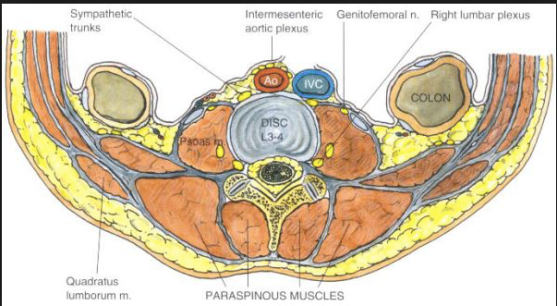
The lumbar sympathetic nerves lie anterolateral to the vertebral body, whereas the somatic nerves lie posterior to the psoas muscle and fascia. The sympathetic chains have anteriorly, aorta on the left and IVC on the right. The aim is to deposit local anesthetic around the nerves from L2 to L4 either with a single injection at L3 or 3 separate injections at L2, L3 and L4. It is used in lower limb ischemia, CRPS, phantom limb, urogenital pain etc

How to do the lumbar sympathetic block?

Patient should be positioned lateral with the side to be blocked up. A point is marked 8 cm from the midpoint of the spinous process of the desired vertebra. Its done under image guidance. A 12 cm 22 G needle is inserted at 45 degree angle directed medially towards the vertebral body which lies at a depth of around 8 cm; if the needle hits the bone at 4-5 cm depth it is likely to be the transverse process and should be redirected cranially or caudally to pass over it. Once the needle hits the vertebral body, it should be redirected slightly antero-laterally till we feels the pop-off of passing the psoas fascia. Then the local anesthetic mixed with radiographic contrast is injected which should form a band around the desired vertebral bodies. If the contrast disappears very quickly, it may be in a vessel. Otherwise, it can go into the psoas muscle or retroperitoneal tissue. Also we should take lateral and anteroposterior images to confirm the correct position.

Complications of lumbar sympathetic block?

Local anesthetic toxicity due to injection into aorta or IVC
Profound motor block or permanent paralysis due to intrathecal injection
Profound hypotension: good iv access and access for resuscitation equipments are a must
Post sympathectomy pain
Ureteric injury, ejaculatory failure
Screen Shot 2019-03-18 at 3.15.22 PMScreen Shot 2019-03-18 at 11.01.44 AM

A FEW FACTS ABOUT COAGULATION FUNCTION, IT’S MONITORING & Regional Anesthesia IN OBSTETRIC PATIENTS

During routine epidural or spinal anaesthesia, accidental puncture of epidural veins occurs in 1–18% of patients

The incidence of hematoma after epidural techniques is estimated to be in the order of 1:150,000 after epidural placement and 1:220,000 after spinal injection in the general population

removal of epidural catheters posed an equal risk to insertion ( Van- dermeulen et al)

Surgery on spinal haematoma should ideally be performed within 8–12 h of the identification of symptoms in order to improve the chances of recovery.

The overall risk of death in those having general anaesthesia for caesarean section was quoted in 2007 as being just over 1:25,000.

The levels of factors VII, VIII and fibrinogen increase and those of anticoagulation factors decrease, causing augmented coagulation and decreased fibrinolysis in pregnancy.

There is no evidence to support routine full blood count (FBC) or coagulation tests in women before the performance of a regional block in those who have had

normal FBC results

no bleeding history

no signs or symptoms of liver disease

no signs or symptoms of pre-eclampsia, abruption or clinical signs of disseminated intravascular coagulation

no recent anticoagulant treatment.

In women with known thrombocytopaenia, a Full Blood Count (FBC) should be checked within 24 h of a regional procedure.

In women with mild to moderate pre-eclampsia, the course of the disease can be unpredictable and so FBC be checked within 6 h. In addition, coagulation tests should be performed if platelets are <100000/mcL or if there is abnormal liver function.

In severe disease, FBC and clotting should be checked immediately before a procedure, as platelet levels in particular can decline rapidly.

Women with pregnancy-induced hypertension alone do not require an FBC before a regional procedure

Activated partial thromboplastin time ratio (APTTR) and international normalised ratio (INR) are slightly decreased in late pregnancy.

In a patient who receives LMWH, if he/she is simultaneously taking NSAID+Aspirin, there is an increased risk if last dose of LMWH is between 12-24 hours; it further increases if last dose is <12 hours

In patients with pre-eclampsia and platelet count between 75000-100000/mcL, there is an increased risk even if coagulation tests are normal; but it increases further if the counts has not been stable (=decreasing platelet count)

#obstetrics , #anesthesia , #coagulation , #anaesthesia

Reference: Abnormalities of Coagulation and Obstetric Anaesthesia, Hilary Swales, AAGBI Core Topics in Anaesthesia 2015

CAUDAL BLOCKS: A FEW FACTS

Most effective for children <20 Kg (~ under 6 years of age) and for dermatomes below T10

Common side effects are weakness of legs, urinary retention etc

Because of this, sometimes a caudal block may necessitate overnight admission

The incidence of epidural hematoma has been reported as 1 in 80000 cases

Dose calculation can be done using Armitage ( 0.5 mL/kg for lumbosacral & 1 mL/kg for lumbar blockade, with 0.25% levobupivacaine ) or Scott formulas

Additives used in caudal block:

Preservative free Ketamine: Extend duration of analgesia; not used in infants <6 months of age due to fear of neurotoxicity

Clonidine : Extend duration of analgesia; not used in preterm infants and neonates due to higher incidence of bradycardia and apnoea. Provides postoperative sedation also.

Opioids when used as additives produce side effects like respiratory depression, pruritus & PONV

#EpiduralBlock , #Anaesthesia , #Anesthesia

References: De Beer DAH, Thomas ML. Caudal additives in children: solutions or problems? Br J Anaesth. 2003; 90: 487–498. Patel D. Epidural analgesia for children. Contin Educ Anaesth Crit Care Pain. 2006; 6(2): 63–66.