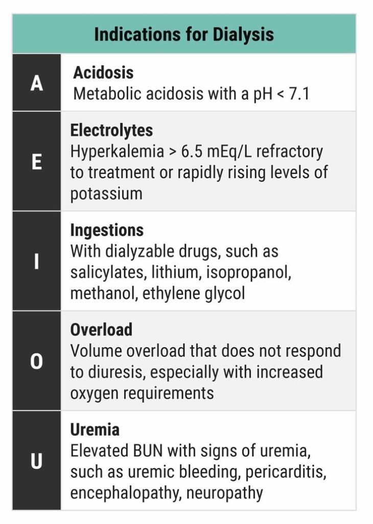
Complications of Laparoscopy

  • The commonest structure that can be injured by the laparoscope is the distended stomach. So pass a nasogastric tube and aspirate the stomach contents especially if the induction has involved prolonged bag and mask ventilation.
  • Regurgitation of gastric contents can happen
  • Pulmonary oedema from fluid infusions and the head-down position
  • Insufflation is the most hazardous phase; A demonstrable gas embolism can occur in 1 out of 2000 patients; so watch for high insufflating pressures and low flow. Signs of gas embolism include arrhythmia, hypotension, cyanosis and cardiac arrest. The safest technique is to use CO2 for the pneumoperitoneum rather than N2O, because CO2 is more soluble and if an embolism occurs, it will resolve faster. Watch the indicators on the insufflating machine continuously during inflation. Pressure over 3 kPa or total volume insufflated exceeding 5 litres are hazardous. Caval compression and reduced venous return, with lowered cardiac output, may be a consequence of intra-abdominal pressure exceeding 4 kPa.
  • The pressure effect of the insufflating gas will also splint the diaphragm and impede the mechanism of breathing.
  • Avoid excessive head down tilt, and always be prepared for laparotomy.
  • The end-tidal CO2 will rise during the course of a prolonged procedure and minute volume should be adjusted to compensate.
  • Pneumothorax and surgical emphysema have been described, associated with prolonged surgery.
  • Shoulder-tip pain, from diaphragmatic irritation, is a common postoperative problem.

AN APPROACH IN DEALING WITH ACCIDENTAL ARETYNOID DISLOCATION AFTER ENDOTRACHEAL INTUBATION

 Follow up the patient:

If voice is not improving: (Better to call the ENT Surgeon to do this-) Do a laryngoscopy and using any instrument, just give a mild pressure on aretynoid; usually it will fall back to correct position.

If speech is improving, advice VOCAL CORD ADDUCTION EXERCISES

Standing position.. Take a deep inspiration
and stop..and hold the breath.. this closes glottis..now strongly fall over and push against a wall…keep it for a few seconds.. Repeat this a few times.. This can force the aretynoid back to normal position by a stretching force… Usually voice is regained by this after 2 days..

Or lift heavy weights after deep inspiration (not for CAD patients)

Plus continue Speech Therapy

Problem occurs, when Aretynoid dislocates, and nobody attempts to relocate it, and it get fixed in that position..

ANKLE BLOCK : NEEDLE DIRECTIONS

Posterior tibial nerve :
Introduce the needle along the medial aspect of the Achilles tendon, at the level of the cephalic (towards head) border of the medial malleolus. Advance, in an anterior direction, towards the posterior border of the tibia (nerve lies just posterior to the posterior tibial artery).If paraesthesia is felt, inject 3-5ml LA. If not, advance to contact the tibia, withdraw 0.5cm and then inject 5-7ml LA.

Sural nerve :
Introduce the needle along the lateral border of the Achilles tendon at the level of the cephalic border of the lateral malleolus.Advance anteriorly towards the fibula.If parasthesia is felt inject 3-5ml LA. If not, inject 5-7ml LA as the needle is withdrawn. This gives subcutaneous infiltration from the Achilles tendon to the fibula.

Infiltration around the remaining three nerves can be performed from a single site. The needle is inserted 1cm lateral to the tendon of extensor hallucis longis (or just lateral to the anterior tibial artery, if palpable), at the level of the cephalic borders of the malleoli. This tendon is prominent on the dorsum of the foot, during extension of the big toe.

Deep peroneal nerve :
From the position described above, advance the needle posteriorly (i.e. at 90¡ to the skin). Inject 3-5ml LA deep to the fascia, on either side of the anterior tibial artery.

Superficial peroneal nerve :
After blocking the deep peroneal nerve, withdraw the needle to just stay in the skin.
Turn the needle towards the lateral malleolus and inject 5ml LA in a subcutaneous band between the lateral malleolus and the anterior border of the tibia. This should reach all the branches of this nerve.

Saphenous nerve :
Again withdraw the needle to just stay in the skin and turn the needle to point towards the medial malleolus.Infiltrate 5ml LA subcutaneously as the needle is advanced towards the medial malleolus. The great saphenous vein lies in this area, just antero-medial to the medial malleolus, in order to infiltrate around the vein, without causing damage, it may be necessary to make a further skin puncture lateral to the vein.

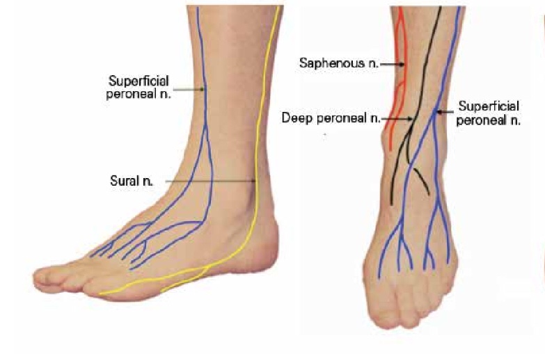
Image courtesy: Semantic Scholar

VIVA AID: IDEAL POSITION OF TIP OF RIGHT IJV CATHETER

  • Different methods have been suggested in the literature
  • Catheter tips positioned approximately 3 cm below the right tracheobronchial angle will lie in close proximity to the atriocaval junction but will remain extracardiac in location
  • Pere P.W. studied correlation between the length of catheter inserted and patient’s height and observed that catheters inserted through right IJV from midcervical point or lower puncture to Height/10cm ended in SVC, while those inserted more than Height/10 + 1cm, 47% ended in right atrium
  • Some studies argued that the catheter tip should lie above the pericardial reflection to prevent serious and potentially lethal complications like cardiac tamponade, malignant arrhythmias, placement in coronary sinus and tricuspid valve damage. The upper limit of the pericardial reflection cannot be seen on a plain chest X-ray, but it is generally accepted to be below the carina.
  • Most clinicians aim to place the catheter tip at the level of the carina with whichever formula they follow. This position carries the risk of migration, thrombosis, and malfunction.
  • A recent paper has demonstrated that a catheter with its tip positioned peripheral to the atriocaval junction was more likely to undergo internal repositioning and venous thrombosis.
  • The lower placement is preferred for left-sided IJV cannulations, because aiming the catheter tip placement above the pericardial reflection in this scenario is more likely to lead to oblique placement and abutment against the wall, which is a risk factor for perforation. It seems, therefore, that a free-floating catheter tip in a wider portion of the IJV is more important than placing the catheter above the pericardial reflection.
  • This becomes even more relevant in the light of the observations by Schuster and colleagues13 that the pericardium can ascend alongside the medial wall of the SVC by up to 5 cm (mean 3 cm). Thus, catheter tip placement even 3 cm above the SVC–RA junction might not obviate the risk of tamponade in all patients.
  • Recently many studies have questioned the placement of the catheter tip in the middle SVC. They argue , emphasizing the importance of having a free-floating tip (not abutting the vessel wall) rather than its placement above the pericardial reflection. This can be achieved by placing the catheter tip in lower SVC and upper RA (target zone, within 2 cm above and 1 cm below the SVC–RA junction). In this position, the SVC is wider, meaning that the catheter tip is likely to float freely with minimal chance of abutment. Furthermore, a catheter placed in this position is amenable to confirmation by TOE.
  • Ahn and colleagues have suggested a radiological landmark-based technique for ‘parking’ the catheter within the target zone. They adjudged the depth of CVC insertion by summing [1] the distance between the sternoclavicular joint and the carina (measured offline from the chest radiograph using an internal measuring tool available on the picture archiving and communication system), [2] the distance between the point of insertion and the sternoclavicular joint, and [3] 1.5 cm. The eventual location of the CVC tip was confirmed by TOE. The authors concluded that the catheter tip was positioned more accurately and the determined depth of CVC insertion correlated better with the actual distance from the skin insertion point to the RA–SVC junction with the radiological landmark-based technique compared with Peres’ formula of central venous catheterization via the right IJV
  • N.B: Also note that from the right internal jugular and left subclavian approaches, the veins respectively take straight and gently curving trajectories to the superior vena cava. However, the right subclavian vein takes a near-right angle turn into the superior vena cava, and the left internal jugular approach incorporates two turns, one into the brachiocephalic vein and a second into the superior vena cava. These turns create potential for the venous side walls to be punctured by a dilator failing to negotiate a curve appropriately.

  • Ref: Quest to determine the ideal position of the central venous catheter tip D. K. Tempe1,* and S. Hasija, British Journal of Anaesthesia 118 (2): 148–50 (2017)

Transurethral resection of the prostate (#TURP) syndrome : A summary 

🚩#TURPsyndrome is diagnosed based on clinical signs, symptoms and biochemical findings
🚩The manifestations are due to hypervolemia, hyponatremia and due to the direct toxicity of the irrigation fluids like 1.5% glycine
▪️FACTORS INCREASING THE ABSORPTION OF THE IRRIGATION FLUID ( AND THUS CONTRIBUTING TO THE HYPERVOLEMIA )
🚩Long duration of the surgery: the irrigation fluid is absorbed at the rate of 20-30 mL/ min and so the volume absorbed increases with the duration of the surgery
🚩High pressure delivery of the irrigation fluid especially from a considerable height; the minimum height required for adequate flow should be used (usually 70 cms)
🚩Low venous pressures
🚩Excessive bleeding (= there are more open veins)
🚩Large prostate (>50g)
▪️CLINICAL FEATURES:
🚩Headache, Restlessness, Agitation, Confusion, Convulsions, Coma; pulmonary oedema may also set in. If patient is under general anesthesia, these symptoms will get masked.
▪️MANAGEMENT FROM SURGICAL SIDE:
🚩Coagulating bleeding points and terminating surgery as soon as possible.
▪️ANESTHETIC MANAGEMENT:
🚩Reduce / stop fluid administration. Diuretics may be required in the presence of pulmonary oedema
🚩Intubation to protect the airway and mechanical ventilation to support respiration may be required
🚩Anti-convulsants, if needed, to treat seizures
🚩Hypertonic saline should be considered for severe hyponatremia (<120 mmol L−1) or in the presence of severe neurological symptoms.

👉🏿N.B.- Central pontine myelinolysis or osmotic demyelinating syndrome (ODS) is more likely to occur with correction of serum Na greater than 8-12 mmol/day and in the setting of chronic hyponatremia (greater than 48 h)
👉🏿Faster rates of administration can potentially lead to central pontine myelinolysis. Treatment should stop once symptoms have resolved or the serum sodium is more than 125 mmol L−1. Such therapy is best delivered in a high-dependency environment.
🔻A template is given below for calculating the volume of hypertonic saline
#anesthesia , #urology ,#hyponatremia , #anaesthesiologist

2016-08-30-09-58-51