- The movement of ions across cell membranes during the depolarisation and repolarisation of myocytes and neurones generates electric potentials.
- Silver metal electrodes covered with a layer of silver chloride gel within an adhesive sponge pad can be used to measure these potentials at the skin.
- Ion movement near the electrode–skin interface induces movement of chloride ions within the gel layer. The ion concentration gradient promotes electron production at the electrode.
- A lead wire and voltmeter attached to the electrode allows measurement of the potential relative to a reference point. The reference point is usually a second skin electrode.
- Signals are then amplified, processed and displayed.
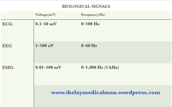
- Skeletal and cardiac muscles have higher amplitudes than cerebral neurones. This is because the amplitude of biological potentials is proportional to the number of simultaneously depolarising cells.
- The frequency of potentials is related to the fluctuating ion activity across cell membranes.
- Skeletal myocytes which undergo tetany have high frequencies of
up to 1 kHz. - Conversely, cardiac myocytes have lower frequencies due to their refractory periods