Cricoarytenoid arthritis: an ominous entity for the anesthesiologist

  • Rheumatoid arthritis is the most common cause of this condition
  • Can also may be associated with bacterial infections, mumps, diphtheria, tuberculosis and ankylosing spondylitis,systemic lupus erythematosus, gout, progressive systemic sclerosis
  • The cricoarytenoid joint has a synovial lining and bursa. Its mobility is vital for speech, respiration, and protection from aspiration.
  • Effusion, pannus formation, joint erosion, and ankylosis may compromise the joint’s functions.
  • Its involvement may be unsuspected or mistaken for asthma until intubation or after extubation and may necessitate a surgical airway.
  • Dysphonia, dyspnea, or stridor should raise suspicion of this possibility.
  • Complete airway obstruction is a well described but an uncommon complication
  • Laryngoscopy may reveal a rough and thick mucosa with narrowing of the vocal chink.
  • Airway obstruction occurs most commonly in patients with long-standing rheumatoid arthritis with polyarticular and systemic involvement
  • But laryngeal stridor has been described as the sole manifestation of this disease too!
  • Always anticipate this as a cause for postoperative stridor in such patients.

COVID 19 AND THE ANAESTHESIOLOGIST: WHAT EXACTLY YOU WILL DO WHEN MANAGING A COVID POSITIVE PATIENT IN OR/ICU OR DURING CPR? A SUMMARY OF 8 SOCIETY GUIDELINES

GENERAL INSTRUCTIONS FOR PERIOPERATIVE SCENARIO (Source: 1 Consensus guidelines for managing the airway in patients with COVID-19 Guidelines from the Difficult Airway Society, the Association of Anaesthetists the Intensive Care Society, the Faculty of Intensive Care Medicine and the Royal College of Anaesthetists, Source: 2 Editorial, anesthesia-analgesia, May 2020,  Source: 3 Anesthesia Patient Safety Foundation and World Federation of Societies of Anesthesiologists, accessed 3/13/2020, Source: 4 Interim guidance for health care providers during covid-19 outbreak from AHA and 5 CDC guidelines)

  • Remember that your personal protection is the priority. Plan ahead as it takes time to apply all the barrier precautions. Before intubation, review and practice donning and doffing the appropriate respiratory protection, gloves, face shield, and clothing.
  • Practice appropriate hand hygiene before and after all procedures.
  • Anaesthesia/ Intubating personnel should don full PPE (Well fitting N95 mask, goggles+ face shield, splash resistant gown, boot covers, double gloves)
  • Patient should wear a mask; continue it during pre-oxygenation
  • When intubated patient being transferred to ICU or transfer from one circuit / ventilator to other, avoid disconnections in patient’s breathing circuit. Put the ventilator on stand by to turn off flows. Clamp ETT with forceps to prevent aerosolization
  • Tracheal intubation of the COVID positive patient is a high risk procedure for the staff, irrespective of the clinical severity of the disease. Do not rush; priority should be to succeed in the first chance. Avoid unreliable, unfamiliar or repeated techniques
  • Know and communicate the plan before entering the room (Use a checklist). Also plan how to communicate once inside the Operating Room (OR) (It will be difficult with the PPE; You may have to use adequate volume while speaking). You can display your plan/algorithm in the OR.
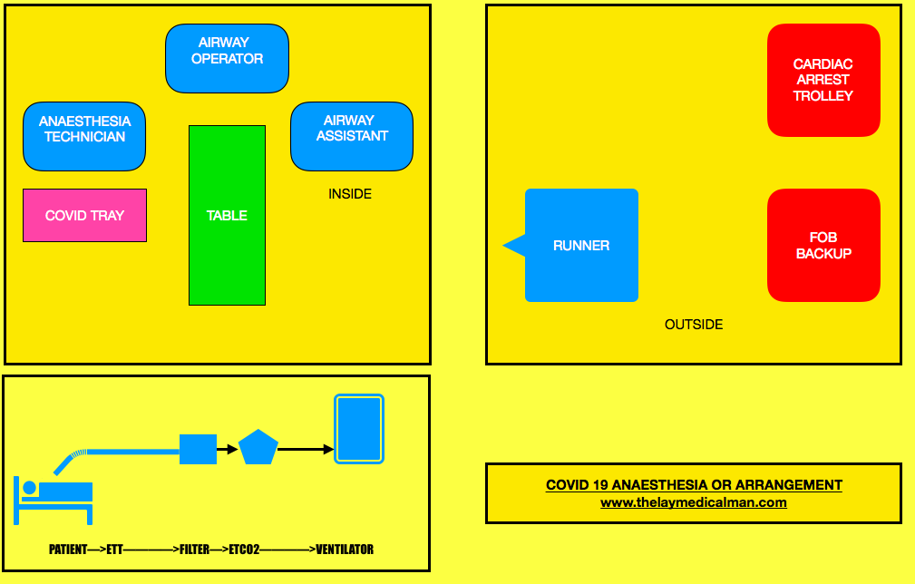
  • Limit staff present at the tracheal intubation: one intubator, one assistant and one to give drugs, equipments and monitor the patient; A runner should be there outside the room. The most experienced anaesthetist available should perform the intubation, to maximise first-pass success. Technician/assistant to keep a distance of 2 m from patient
  • Touch as less as possible once inside the room
  • Ideally we should intubate in a negative pressure room with >12 air changes per hour
  • If this is not available, switch off air conditioner/ positive pressure, 20 minutes before and 20 mins after Aerosol Generating Procedures

PREPARATION OF DRUG/ EQUIPMENT

PROCEDURE:

  • Most of this should happen outside the room
  • Pre-procedure machine check to ensure no leak. Check circuit
  • Create a COVID Intubation Trolley (This can also be used in the ICUs)
  • Arrange N95 mask and 2 HEPA filters to attach between tracheal tube and breathing circuit and between expiratory limb and anaesthesia machine
  • Standard monitors, cannulas, instruments, drugs
  • If patient is on HCQ, it will be better to avoid glycopyrrolate and ondansetron
  • Sterile plastic covers: for protecting monitors, ventilator or anaesthesia machine and for covering the cable covering the laryngoscope handle. Also keep stylet, appropriate size cuffed tube,10 ml syringe for cuff inflation, oral suction catheter etc
  • Face mask
  • Airways
  • A second generation Supra Glottic Airway device for airway rescue
  • Use 5 minutes of preoxygenation with 100% oxygen and RSI techniques to avoid manual ventilation of patient’s lungs and the potential aerosolization of virus from airways.
  • No bag mask manual ventilation. Holding the mask: 2-person, 2-handed, with a VE grip technique (rather than the C- technique) to improve seal. If you are forced to mask ventilate, use a 2-person, low flow, low pressure technique
  • Intubating dose of Rocuronium or Suxamethonium should be given along with propofol or ketamine (can avoid cardiovascular collapse in some situations): Intubate after 90 seconds (prevent coughing)
  • Indirect laryngoscopy with video laryngoscope & intubation under the transparent plastic sheet on the patient
  • Inflate the ETT cuff immediately after tube placement, before starting ventilation
  • HEPA shield antiviral filter connected to ETT & then connect ETT to ventilator breathing circuit
  • Avoid all Aerosol Generating Procedures (AGP) like high flow nasal oxygen, NIV, bronchoscopy and tracheal suction without a closed suction facility. Routine use of supraglottic airway devices unless in unanticipated difficult airway should be avoided
  • Use a closed suction system
  • Confirm correct position of the tracheal tube. Confirmation of the ETT position will be difficult while wearing PPE; so for this purpose we may have to rely on inspection of bilateral chest examination, observation of ETCO2 waveforms etc.
  • Have a vasopressor ready for managing hypotension if it happen post-intubation
  • Push-twist all connections to avoid circuit disconnections
  • Clamp tube and pause ventilator for all airway manoeuvres and for attempting to resolve circuit disconnections
  • Place a nasogastric tube if necessary.
  • If COVID-19 status has not been confirmed, take a deep tracheal aspirate using closed suction
  • Institute mechanical ventilation and stabilize patient, as appropriate.
  • Lung protective ventilation strategies: Small TV:6 ml/Kg [Predicted body wt= Ht in cm-100 (males) & Ht- 110 (females)]. Plateau pressure </= 30 cm H2O. PEEP= 10-15 mm Hg, Adjust FiO2 to achieve reasonable PaO2 (>60 mm Hg). Target SaO2 88-95%. pH >/= 7.25 (Permissive Hypercapnia)
  • Use only metered dose inhalers if bronchodilators indicated at any point ( avoid nebulisation)
  • Use of intravenous anesthesia would be preferred to the use of a volatile gas anesthetic machine in the ICU environment, especially given that many of these patients are not going to be rapidly recovered and extubated following the procedure. (Source:6)
  • Rule out pneumothorax if there is difficulty in ventilation ( Lung USG, CXR)
  • Clean the room 20 minutes after tracheal intubation or any AGP.
  • Use low gas flows and closed circuits
  • Prophylactic anti emetic before extubation
  • Adequate Pain management: Morphine/Fentanyl boluses
  • All efforts to prevent coughing including lidocaine/dexmedetomidine
  • To prevent aerosol generation extubation also should be performed under transparent sheet
  • O2 by nasal cannula / face mask. When wearing nasal prongs, a surgical mask can be worn by the patient over the prongs to reduce droplet spread. Should higher oxygen requirements necessitate use of a mask, non-rebreather masks with an attached exhalation filter can be used.
  • NIV or High flow O2 can cause aerosol generation: so better to avoid
  • Ensure the availability of ambu bag with filter during transfer
  • After leaving the room, do a meticulous doffing of PPE
  • In the ICU: The use of CPAP/BiPAP may increase the risk of delayed deterioration leading to need for emergent intubation and increased risk of mistakes in donning PPE due to time pressures to resuscitate. In general, CPAP/BiPAP should be avoided in patients with Covid 19 and should never be used outside of appropriate airborne/droplet isolation.
  • All airway equipment must be decontaminated and disinfected according to appropriate hospital policies.
  • After removing protective equipment, avoid touching hair or face before washing hands.
  • NB: N95 mask disinfection: Either cycle through 4 masks in series: one for each day, then repeat (OR heat the mask to 70 degree for 30 minutes (UV light, alcohol, bleach and touching the metal of the oven during heating…all these can degrade the mask)

EMERGENCY INTUBATION IN THE CRITICAL CARE UNIT (Source: 6 Wax, R.S., Christian, M.D. Practical recommendations for critical care and anesthesiology teams caring for novel coronavirus (2019-nCoV) patients. Can J Anesth/J Can Anesth(2020))

  • Here also the same principles cited above should apply; additional points are
  • Higher level of precautions are needed here as there is high-level viral shedding due to severity of patient illness and procedures associated with resuscitation or intubation may generate aerosols
  • All personnel in the room must be using appropriate PPE, including either a fit-tested N95 mask or a PAPR. The procedure should be attempted by the most skilled person. Recurrent traffic of people bringing equipment into the room may increase the risk of viral transmission. 
  • Clinicians should strongly consider pneumothorax in any ventilated patient with sudden respiratory deterioration. Portable ultrasound may be used to quickly assist in the diagnosis of a pneumothorax, as arranging for a CXR will lead to delay in intervention.

DIFFICULT AIRWAY

Compared to the normal patient, after the first failure of intubation itself, we should order for Front Of Neck Access (FONA) set. And in the next step, we can either do step B (SGD) or step C ( Facemask). Because of this, we will move fast towards the final step of FONA in the Covid difficult airway algorithm.

DIFFICULT AIRWAY: NORMAL PATIENT VS COVID PATIENT

Prevention and management of respiratory or cardiac arrest: Protected Code Blue (PCB) (Source: 7 Resuscitation Council. Resuscitation Council UK Statement on COVID-19 in relation to CPR and resuscitation in healthcare settings. 2020. Source: 8 Wax, R.S., Christian, M.D. Practical recommendations for critical care and anesthesiology teams caring for novel coronavirus (2019-nCoV) patients. Can J Anesth/J Can Anesth(2020))

  • Do not listen or feel for breathing by placing your ear and cheek close to the patient’s mouth
  • Full Aerosal Generating Procedure (AGP) Personal Protective Equipment (PPE) must be worn by all members of the resuscitation/emergency team before entering the room.
  • Sets of AGP PPE must be readily available where resuscitation equipment is being locally stored.
  • No chest compressions or airway procedures such as those detailed below should be undertaken without full AGP PPE.
  • Once suitably clothed, start compression-only CPR and monitor the patient’s cardiac arrest rhythm as soon as possible.
  • Do not do mouth-to-mouth ventilation or use a pocket mask. If the patient is already receiving supplemental oxygen therapy using a face mask, leave the mask on the patient’s face during chest compressions as this may limit aerosol spread.
  • If not in situ, but one is readily available, put a simple oxygen mask on the patient’s face. Restrict the number of staff in the room (if a single room). Allocate a gatekeeper to do this. 
  • Tracheal intubation or SGA insertion must only be attempted by individuals who are experienced and competent in this procedure. 
  • Dispose of, or clean, all equipment used during CPR following the manufacturer’s recommendations and local guidelines.
  • Any work surfaces used for airway/resuscitation equipment will also need to be cleaned according to local guidelines.
  • Specifically, ensure equipment used in airway interventions (e.g. laryngoscopes, face masks) is not left lying on the patient’s pillow, but is instead placed in a tray.
  • Do not leave the Yankauer sucker placed under the patient’s pillow; instead, put the contaminated end of the Yankauer inside a disposable glove. 

Lower risk resuscitation interventions:

Placement of an oral airway
Placement of an oxygen mask with exhalation filter on patient (if available)
Chest compressions
Defibrillation, cardioversion, transcutaneous pacing
Obtaining intravenous or intraosseous access
Administration of intravenous resuscitation drugs

Higher risk resuscitation interventions more likely to generate aerosol and/or increase risk of viral transmission to staff

Bag-mask ventilation
CPAP/BiPAP
Endotracheal intubation/surgical airway
Bronchoscopy
Gastrointestinal endoscopy

MEDIASTENAL TUMOURS & THE ANESTHESIOLOGIST: SPECIFIC POINTS

  • A preoperative CT scan will show the site, severity, and extent of the airway compromise to assess the level and degree of obstruction.
  • Assess the vocal cord function preoperatively
  • Lung function tests to look for the extent of intrathoracic or extrathoracic obstruction.
  • ECHO to rule out pericardial effusion and cardiac compression.
  • Premedication with benzodiazepine is generally avoided if there is risk of airway compromise.
  • Airway equipment—rigid bronchoscopy and difficult airway trolley, jet ventilation, cardiopulmonary bypass (CPB) should be there as standby. Femoro femoral bypass is the most common setup.
  • COMPLICATIONS DUE TO MASS EFFECT OF THE TUMOUR:
  • Vascular compromise—SVC Obstruction ( SVCO ) and pulmonary vessel obstruction
  • Laryngeal nerve palsy
  • Dysphagia
  • STRIDOR and airway compromise may be an important symptom
  • Inspiratory stridor (laryngeal)—obstruction above the level of glottis
  • Expiratory stridor (tracheobronchial)—obstruction in the intrathoracic airways
  • Biphasic stridor—obstruction between glottis and subglottis or a critical obstruction at any level
  • Sometimes you may have to go for a microlaryngoscopy tube (MLT)
  • TAKE CARE:
  • Aim to avoid worsening of cardiac compression, airway occlusion, and SVC obstruction.
  • IV cannula in the lower extremity
  • Induction in sitting position (semi Fowler’s position)
  • Inhalational (preferred choice) or IV induction agent titrated to effect
  • Choose spontaneous ventilation with LMA
  • Awake fibreoptic technique if intubation is necessary with a reinforced smaller calibre and longer endotracheal tube
  • Postoperative airway obstruction due to airway oedema, tracheomalacia, and bleeding warrant the need for awake extubation in ITU. The following steps would aid in an uneventful extubation:
  • Test for leak around the endotracheal tube cuff.
  • Administer dexamethasone or chemo radiotherapy in sensitive tumours to shrink size of tumour.
  • Use adrenaline nebulisers.
  • Extubate over airway exchange catheters.
  • SVCO: challenges during anaesthesia
  • Need for supplemental oxygen
  • Orthopnoea—induction in the sitting-up position
  • IV cannula in the lower extremity
  • Airway oedema
  • Mucosal bleeding
  • Laryngeal nerve palsy
  • Haemodynamic instability due to decreased venous return
  • OTHER CONCERNS
  • General anaesthesia, causes loss of intrinsic muscle tone, decreased lung volumes, and decreased transpleural pressure gradient
  • Positive pressure ventilation, can precipitate severe hypotension and also increases intrathoracic tracheal compression
  • Coughing, as it can cause complete airway obstruction by positive pleural pressure, increasing intrathoracic tracheal compression
  • Following gas induction, the patient stops breathing and if you are unable to ventilate her: Follow difficult or failed intubation guidelines. But cricoid puncture and emergency tracheostomy are futile if the level of airway obstruction is at the intrathoracic tracheobronchial tree: Try a change in position—lateral, sitting up, or prone—to decrease the mechanical effect of the tumour. Avoid positive pressure ventilation for fear of luminal closure. Low-frequency jet ventilation with Sander’s injector or high-frequency translaryngeal jet ventilation with Hunsaker’s catheter is one option. CPB bypass and ECMO to restore oxygenation when other measures fail.
  • Following chemotherapy in ICU, if patient develops hyperkalemia, Tumour Lysis Syndrome should be there in the differential diagnosis
  • ALSO NOTE
  • During inspiration, the intrathoracic airways expand along with the expanding lungs. In contrast, the extrathoracic airways diminish in caliber during inspiration due to their intraluminal pressure being lower than the atmospheric pressure. The reverse happens during expiration.
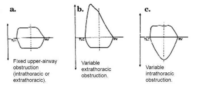
  • Flow volume loop in upper-airway obstruction:
  • Fixed lesions [extrathoracic or intrathoracic] are characterized by lack of changes in caliber during inhalation or exhalation and produce a constant degree of airflow limitation during the entire respiratory cycle. Its presence results in similar flattening of both the inspiratory and expiratory portions of the flow-volume loop
  • Variable lesions are characterized by changes in airway lesion caliber during breathing. Depending on their location (intrathoracic or extrathoracic), they tend to behave differently during inhalation and exhalation.
  • In the case of an extrathoracic obstructing lesion, during inspiration, there is acceleration of airflow from the atmosphere toward the lungs, and the intraluminal pressure decreases with respect to the atmospheric pressure due to a Bernoulli effect, resulting in the limitation of inspiratory flow seen as a flattening in the inspiratory limb of the flow-volume loop. During expiration, the air is forced out of the lungs through a narrowed (but potentially expandable) extrathoracic airway. Therefore, the maximal expiratory flow-volume curve is usually normal.
  • Variable intrathoracic constrictions expand during inspiration, causing an increase in airway lumen and resulting in a normal-appearing inspiratory limb of the flow-volume loop. During expiration, compression by increasing pleural pressures leads to a decrease in the size of the airway lumen at the site of intrathoracic obstruction, producing a flattening of the expiratory limb of the flow-volume loop
  • Screen Shot 2018-09-19 at 1.34.27 AM

VOCAL CORD PALSIES

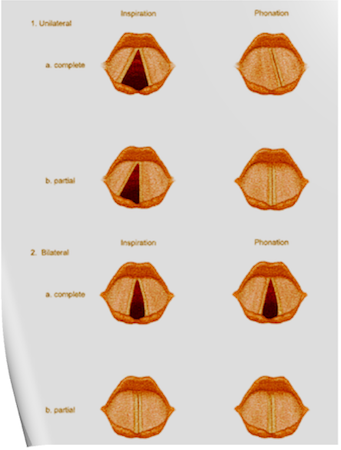
Under normal circumstances, the vocal cords meet in the midline during phonation. On inspiration, they move away from each other. They return toward the midline on expiration, leaving a small opening between them. When laryngeal spasm occurs, both true and false vocal cords lie tightly in the midline opposite each other.

The recurrent laryngeal nerve (RLN) carries both abductor and adductor fibers to the vocal cords.

Selmon’s law: The abductor fibers are more vulnerable, and moderate trauma causes a pure abductor paralysis. Severe trauma causes both abductor and adductor fibers to be affected. N.B.:- Pure adductor paralysis does not occur as a clinical entity.

Scenario 1- PURE UNILATERAL ABDUCTOR PALSY: As adduction is still possible on the affected side, the opposite cord come and meet in the midline on phonation. However, only the normal cord abducts during inspiration.

Scenario 2- COMPLETE UNILATERAL PALSY OF THE RLN: Both abductors and adductors are affected. On phonation, the unaffected cord crosses the midline to meet its paralyzed counterpart, appearing to lie in front of the affected cord. On inspiration, the unaffected cord moves to full abduction.

Scenario 3- BILATERAL INCOMPLETE ABDUCTOR PALSY: When there is incomplete bilateral damage to the recurrent laryngeal nerve, the adductor fibers draw the cords toward each other, and the glottic opening is reduced to a slit, resulting in severe respiratory distress.

Scenario 4- COMPLETE BILATERAL PALSY OF THE RLN: With a complete palsy, each vocal cord lies midway between abduction and adduction, and a reasonable glottic opening exists.

Thus, bilateral incomplete palsy is more dangerous than the complete variety.

Scenario 5- DAMAGE TO SUPERIOR LARYNGEAL NERVE/S: Damage to the external branch of the superior laryngeal nerve or to the superior laryngeal nerve trunk causes paralysis of the cricothyroid muscle (the tuning fork of the larynx), resulting in hoarseness that improves with time because of increased compensatory action of the opposite muscle. The glottic chink appears oblique during phonation. The aryepiglottic fold on the affected side appears shortened, and the one on the normal side is lengthened. The cords may appear wavy. The symptoms include frequent throat clearing and difficulty in raising the vocal pitch.

Scenario 6- TOTAL BILATERAL PARALYSIS OF VAGUS NERVES: This affects the recurrent laryngeal nerves and the superior laryngeal nerves. In this condition, the cords assume the abducted, cadaveric position. The vocal cords are relaxed and appear wavy. A similar picture may be seen after the use of muscle relaxants.

Screen Shot 2018-06-26 at 11.01.24 PM

N.B:- Topical anesthesia of the larynx may affect the fibers of the external branch of the superior laryngeal nerve and paralyze the cricothyroid muscle, signified by a “gruff” voice. Similarly, a superior laryngeal nerve block may affect the cricothyroid muscle in the same manner as surgical trauma does.

Reference: Benumof and Hagberg’s Airway Management, Third edition

RESPIRATORY PHYSIOLOGY IN THE ELDERLY 👴🏻

🍃Ventilatory responses to hypoxia and hypercapnia are impaired secondary to reduced central nervous system activity.

🍃The respiratory depressant effects of benzodiazepines, opioids, and volatile anesthetics are exaggerated.

🍃These changes compromise the usual protective responses against hypoxemia after anesthesia and surgery in elderly patients.

🍃The loss of elastic recoil combined with altered surfactant production leads to an increase in lung compliance.

🍃Increased compliance leads to limited maximal expiratory flow and a decreased ventilatory response to exercise.

🍃Loss of elastic elements within the lung is associated with enlargement of the respiratory bronchioles and alveolar ducts, and a tendency for early collapse of the small airways on exhalation.

🍃There also is a progressive loss of alveolar surface area secondary to increases in size of the interalveolar pores of Kohn. This results in increased anatomic dead space, decreased diffusing capacity, and increased closing capacity all leading to impaired gas exchange.

🍃Loss of height and calcification of the vertebral column and rib cage lead to a typical barrel chest appearance with diaphragmatic flattening.

🍃The flattened diaphragm is mechanically less efficient, and function is impaired further by a significant loss of muscle mass associated with aging. Functionally, the chest wall becomes less compliant, and work of breathing is increased.

🍃Total lung capacity is relatively unchanged.

🍃Residual volume increases by 5% to 10% per decade.

🍃Vital capacity decreases.

🍃Closing capacity increases with age.

🍃Functional residual capacity (FRC) is determined by the balance between the inward recoil of the lungs and the outward recoil of the chest wall. FRC increases by 1%–3% per decade because at relaxed end expiration, the rate of decrease in lung recoil with aging exceeds that of the rate of increase in chest wall stiffness.

🍃In younger individuals, closing capacity is below functional residual capacity. At 44 years of age, closing capacity equals functional residual capacity in the supine position, and at 66 years of age, closing capacity equals functional residual capacity in the upright position.

🍃When closing capacity encroaches on tidal breathing, ventilation-perfusion mismatch occurs.

🍃When functional residual capacity is below closing capacity, shunt increases, and arterial oxygenation decreases. This results in impairment of preoxygenation. Increased closing capacity in concert with depletion of muscle mass causes a progressive decrease in forced expiratory volume in 1 second by 6% to 8% per decade.

🍃Increases in pulmonary vascular resistance and pulmonary arterial pressure occur with age and may be secondary to decreases in cross-sectional area of the pulmonary capillary bed. Hypoxic pulmonary vasoconstriction is blunted in elderly individuals and may cause difficulty with one-lung ventilation.

Ref: Geriatric Anesthesia 2/e , Miller’s Anesthesia 7/e

#Physiology , #Anesthesia , #Geriatrics