AEQ 2: Pulmonary Function Tests (PFT)

  • Anaesthesia Exam Question 2
  • PFTs are done to evaluate the mechanical and gas exchange functions of the lung; most important of these include :
  • Spirometry
  • Lung Volumes & Elasticity
  • Gas Exchange Function Tests
  • SPIROMETRY
  • It’s the measurement of dynamic lung volumes during forced expiration and inspiration. It includes
  • Forced Vital Capacity (FVC)
  • Forced Expiratory Volume in one second (FEV1) &
  • Maximum expiratory flow during the middle 50% (25-75%) of the vital capacity- this is a sensitive index of small airway obstruction 
  • The severity of COPD is graded based on the FEV1 values:
  • Mild COPD- FEV1 >70% of predicted
  • Moderate COPD- FEV1 50-69% of predicted
  • Severe COPD- FEV1 <50% of predicted
  • In COPD and asthma, the FEV1 will be reduced more markedly than FVC resulting in a reduction in FEV1/FVC ratio (>70%)
  • The response to bronchodilators in the form of an increase in FEV1 and FVC indicates reversibility and helps in diagnosis- a response >15% from the baseline value shows significant reversibility and points towards a diagnosis of bronchial asthma
  • TLC and RV will be increased in COPD
  • THE FLOW VOLUME(FV) LOOPS
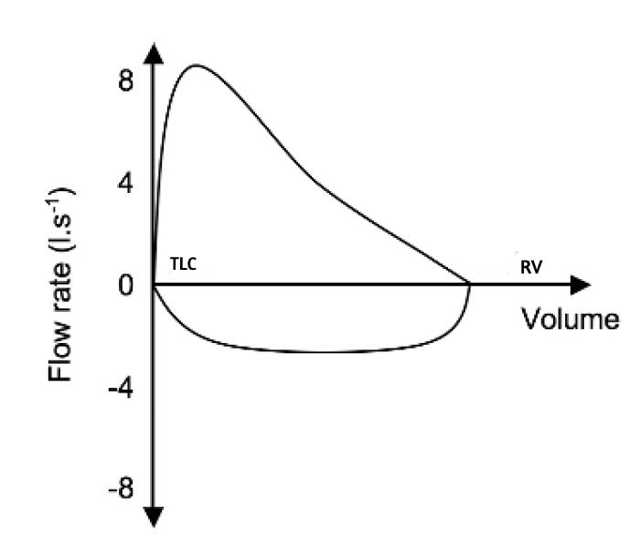
  • The normal FV loop:
  • The patient takes a vital capacity breath and starts the test with a forced expiration
  • The loop rises rapidly to a flow rate of 8-10 l/sec followed by a steady decrease as the expiration continue from left to right- the patient will be unable to cross the maximal flow line because of the dynamic compression of the airways. So the normal loop represents the maximum flow rate at each lung volume
  • Then inspiration occurs from RV to TLC in a right to left direction to achieve a flow rate of 4-6 l/sec and has a squarer shape
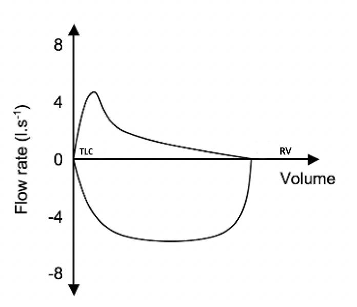
  • FV loop: COPD
  • COPD reduces flow rates all through the expiration with increased concavity or a ‘scooped out’ appearance of the expiratory limb. PEFR is also reduced
  • Due to gas trapping, the RV (and TLC) is increased. The inspiratory flow rates are only slightly reduced
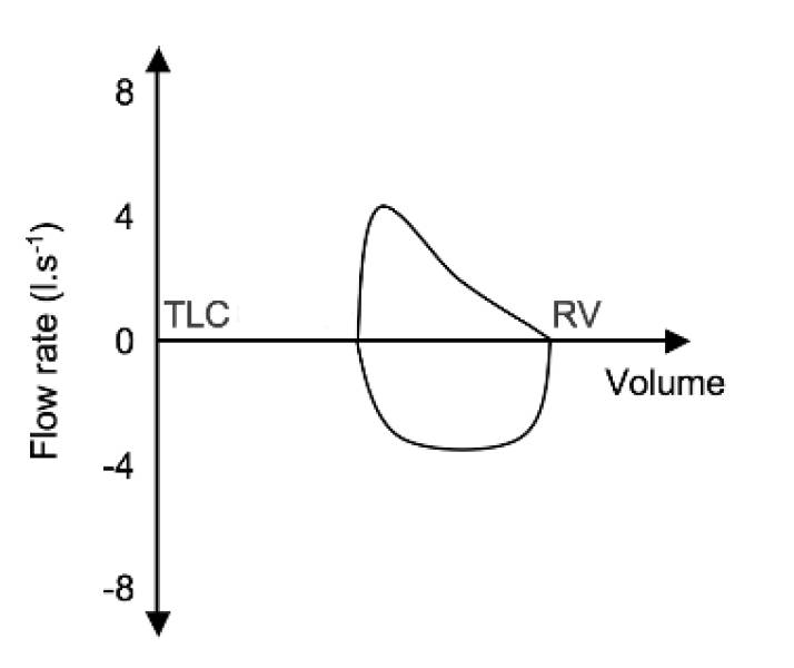
  • FV loop: Restrictive Diseases
  • Restrictive diseases shifts the left hand side of the curve towards right because of a marked reduction in TLC while preserving RV. PEFR is also reduced.
  • FV loop: Variable Extrathoracic Obstruction – Problem: during inspiration
  • e.g. Laryngomalacia, vocal cord paralysis, subglottic stenosis, goitres, tracheomalacia of extrathoracic trachea
  • The negative pressure generated in the airway during inspiration makes the airway collapse thus markedly reducing the inspiratory flow, but retains the square shape
  • The positive pressure generated during expiration, keeps the airway open. So the expiratory limb may be near normal
  • The TLC and RV are generally unaffected.
  • FV Loop: Variable Intrathoracic Obstruction
  • e.g. Tracheomalacia of intrathoracic trachea and tracheal tumours
  • If the obstruction is intrathoracic, the negative pressure generated in the intrapleural compartment during inspiration will help to pull and open up the airways. So the inspiratory limb will be near normal. 
  • The positive pressure generated during expiration will make the airway further narrower. So the expiratory flow will be markedly reduced, similar to that happens in COPD.
  • The TLC and RV are generally unaffected.
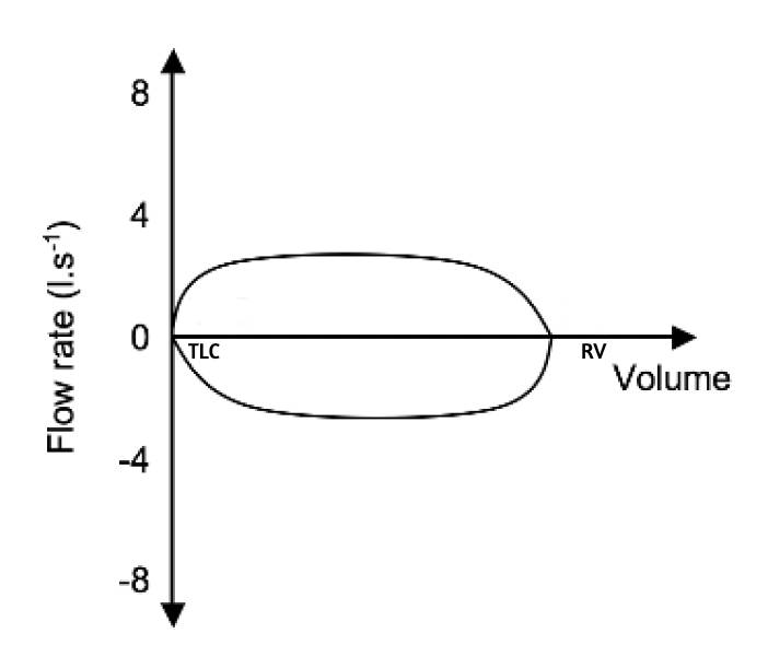
  • FV Loop: Fixed Large Airway Obstruction
  • e.g. Tracheal stenosis
  • Here the peak expiratory and inspiratory flows are determined by the diameter of the orifice- so both the expiratory and inspiratory limbs show markedly reduced flow.
  • The TLC and RV are generally unaffected.
  • GAS EXCHANGE FUNCTION TESTS
  • TLCO measures the integrity and functioning of the gas exchange surface of the lung. This will be reduced in emphysema and is also a good indicator of the severity of the disease. PEFR is also reduced.
  • Arterial Blood Gas analysis and Exercise testing are additional tests that will reflect the functioning of lung function

VIVA SCENE: C-SPINE X-RAY

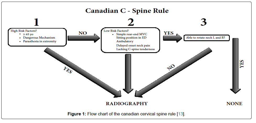
WHETHER TO DO C SPINE IMAGING IN TBI; CRITERIAS:

1. Screen Shot 2019-08-11 at 9.23.39 pm

2. Under the NEXUS guidelines, when an acute blunt force injury is present, a cervical spine is deemed to not need radiological imaging if all the following criteria are met:

  • There is no posterior midline cervical tenderness
  • There is no evidence of intoxication
  • The patient is alert and oriented to person, place, time, and event
  • There is no focal neurological deficit (see focal neurological signs)
  • There are no painful distracting injuries (e.g., long bone fracture)

Screen Shot 2019-08-26 at 11.12.53 am

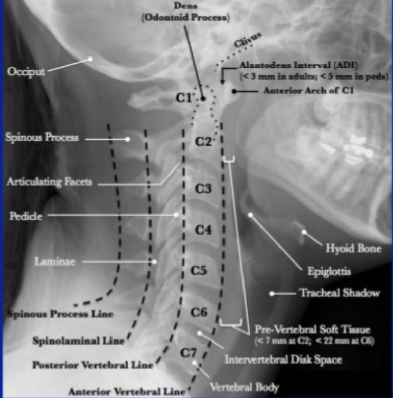
Lateral C-Spine Radiograph. (AABCDs)

A—Adequacy: An adequate film should include all seven cervical vertebrae and C7/T1 junction with optimum density so that the soft tissue shadow is visible clearly.

A—Alignment

a. Atlanto occipital alignment: The anterior and posterior margins of the foramen magnum should line up with the dens and the C1 spinolaminar line.

b. Vertebral alignment

Look for the following four lines (any incongruity= should be considered as evidence of ligamentous injury or occult fracture)

1. Anterior vertebral line—joining the anterior margin of vertebral bodies

2. Posterior vertebral line—joining the posterior margin of vertebral bodies

3. Spinolaminar line—joining the posterior margin of spinal canal

4. Spinous process/ Interspinous line—joining the tips of the spinous processes

B Bony Landmark: vertebral bodies, pedicles, laminae, and the facet joints are inspected

C Cartilagenous space: Predental space or the Atlanto-Dental Interval (ADI): which is the distance from dens to the body of C1. ADI should be < 3 mm in adults and < 5 mm in children. An increase in ADI depicts a fracture of the odontoid process or disruption of the transverse ligament

D—Disc space

Disc spaces should be roughly equal in height and symmetrical.

Loss of disc height can happen in degenerative diseases.

S—soft tissue

Prevertebral soft tissue space thickness can help in the diagnosis of retropharyngeal haemorrhage, which can be secondary to vertebral fractures. Maximum allowable distances :

Nasopharyngeal space (C1): 10 mm

Retropharyngeal space (C2–C4): 5–7 mm

Retrotracheal space (C5–C7): 14 mm in children and 22 mm in adults

VIVA SCENE IMAGING: CHEST X-RAY (CXR)

  • Helpful MNEMONICS: ‘PRIP ABCDEFGHI’. Also in inspiratory films the level of the diaphragm is at the level of ribs 5/6 anteriorly and 8/10 posteriorly. (AR6PR10). 
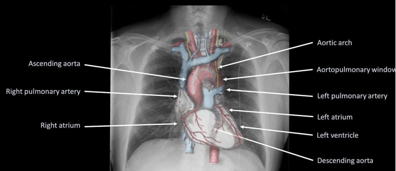
  • Right heart border is formed predominantly by the right atrium along with the lower part of SVC, whilst the left border is formed by the aortic arch, pulmonary artery, left atrium, and ventricle. The right and left ventricle forms the inferior border. The right diaphragm is normally higher than the left due to the liver.
  • Areas where pathology is commonly missed: Apices (including behind the 1st rib and clavicle)—small pneumothoraces and masses. Hila—masses and lymph nodes; left hilum is 1–2 cm higher than right. Behind the heart—left lower lobar collapse and hiatus hernia. Below the diaphragm—free gas. Soft tissues—breast shadow or absence (look for lung and bone metastasis)
  • This is CXR of Mr Roy (Patient identity) done on 14/08/2019 (date of study)
  • Its an AP film (Projection)
  • Non rotated (Rotation)
  • Expiratory film (Inspiratory/Expiratory)
  • Adequately penetrated (Penetrated)
  • Trachea is central, no foreign bodies or other abnormalities (A– Airway)
  • On both sides, the bones and soft tissues appear normal; ribs, sternum, scapula, clavicle, spine, and humerus has no deposits or fractures (B-Bones and soft tissues)
  • The cardiac silhoutte is normal (C-Cardiac silhoutte)
  • There is no free air under the diaphragm. Bilateral costo- and cardio-phrenic angles are clear (D– Diaphragm and angles)
  • No effusions. Pleura is not visible (E-Effusion)
  • Both sides, the lung fields appear normal (F-Fields = lung fields)
  • Gastric bubble visible at left upper abdomen (G=Gastric bubble)
  • The hila and the mediastenum appears to be normal and not displaced. The CT ratio is less than 50% (H-Hila and mediastenum)
  • Endotracheal tube tip is seen 5 cms above the carina or T4/T5 interspace. NG tube passed down midline, past level of diaphragm, and deviates to left with tip seen in stomach. ECG leads and electrodes are noted (I=Insertions, Interventions: tubes and lines, chest drains, pacemakers, and metallic valves and artefacts)
Screen Shot 2019-08-25 at 5.10.08 pm
  • If an intrathoracic opacity is in anatomical contact with the heart border, then the opacity will obscure that border. If an intrathoracic opacity is in the posterior pleural cavity so not in direct anatomical contact with the heart border, this causes an overlap but not an obliteration of that border (Silhoutte sign)
  • When the alveoli no longer contain air and opacify, the air-filled bronchi passing through the alveoli may be visible as branching linear lucencies. (Air bronchogram)
  • Linear opacities measuring 1–6 cm extending from periphery to the hila caused by distension of anastomotic channels between peripheral and central lymphatics.(Kerley A lines)
  • Short horizontal lines, due to oedema of the interlobular septae, situated perpendicularly to the pleural surface at the lung base (Kerley B lines)
  • Reticular opacities at the lung base (Kerley C lines)
  • The mediastinal masses can be identified on the PA views but lateral films and/or CT scans are required to confirm further diagnosis
  • Anterior mediastenum: Anterior to trachea, Middle mediastenum: Anterior to heart, Posterior mediastenum: Posterior to heart
  • ANTERIOR MEDIASTENAL MASS: (4 T’s) Thymic tumour -Teratoma- Thyroid –  Terrible lymph nodes (LN) Ascending aortic aneurysm, MIDDLE MEDIASTENAL MASS (BAL) Bronchogenic cyst – Arch aneurysm- LNs, POSTERIOR MEDIASTENAL MASS: as expected it can be from Spine/Neurogenic; also hiatus hernia
  • Hilar mass: Most commonly TB/Sarcoidosis; also lymphoma
  • Collapse: Triangular opacity, Crowding of ribs, Mediastenal displacement, hyperinflation of other lobes
  • Consolidation: confluent ill defined opacity, bat wing distribution, no volume loss, air bronchogram
  • Effusion: Erect: obliteration of the costo- and cardio-phrenic angles and opacity with meniscus. Supine: graded haze giving a ground glass opacity
  • Emphysema: Decreased lung markings, flat diaphragm and hyperexpanded lung, Presence of bullae and peribronchial thickening, signs of cor pulmonale, right ventricular enlargement, pulmonary hypertension—enlargement of the central pulmonary arteries with oligaemic peripheral lung fields

A FEW CLUES IN INTERPRETING AN ISOLATED PROLONGATION OF ACTIVATED PARTIAL THROMBOPLASTIN TIME (aPTT)

aPTT tests the intrinsic and common pathways of coagulation

Though it is included commonly as a part of coagulation profile assessment, it’s primary uses are to detect coagulation factor deficiency and titration of heparin therapy

An isolated elevation of aPTT may indicate

deficiency of Factor VIII or IX or XI or XII

acquired clotting factor inhibitors

presence of Lupus anticoagulant

N.B.:- Factor VIII deficiency is Haemophilia A, Factor IX deficiency is Haemophilia B and Factor XI deficiency is Haemophilia C

If factor levels are >30% of normal, aPTT may remain normal, for e.g. in mild von Willebrand disease [raised aPTT + prolonged Bleeding Time (BT)], in mild hemophilia etc

Reference: Martlew V. Peri-operative management of patients with coagulation disorders. Br J Anaesth. 2000; 85(3): 446–455.

PROJECT THE RAYS CORRECTLY: Basic facts about projection in X ray films

A postero-anterior (PA) projectionn will not produce as much magnification of the heart and mediastinum as an antero-posterior (AP) projection. So PA films are the preferred ones. Here the scapula is rotated; so the lung fields are clear.

A PA film is taken with the film cassette in front of the patient and the beam delivered from behind with the patient in an upright position.

Portable films and those taken in ICUs are all AP projection. All anterior structures appear magnified—heart, mediastinum, sternum, clavicles, and ribs

The supine position causes distension of the upper lobe blood vessels, which may be confused with elevated left atrial pressure

A lateral X-ray is useful in viewing retrosternal and chest wall lesions, localising lesions in the AP dimension, locate lesions behind the left side of the heart or in the posterior recesses of the lungs. Lateral decubitus—used in diagnosing very small collection of air or fluid in the pleural space.

A left lateral (with the left side of the chest against the film and the beam projected from the right) is the standard projection.

The heart is magnified less with a left lateral as it is closer to the film.

To visualize lesions in the left hemithorax, obtain a left lateral film and for right-sided lesions a right lateral.

Expiratory films are used to assess air trapping in bronchial obstruction such as a foreign body.

A pneumothorax always appears larger on an expiratory film and occasionally a small pneumothorax may only be visible on expiration.

Films if accidentally taken in expiration, can result in spurious magnifcation of the heart and mediastinum.

  • PROJECTION: The different projections of importance to us are Postero Anterior (PA), Antero Posterior (AP), Lateral, Supine, and Lateral decubitus.
  • ROTATION: An image is not rotated if the clavicular heads are equidistant to the
    corresponding thoracic spine
  • INSPIRATION: An inspiratory picture shows a ‘lot of lung’. In inspiratory films the
    level of the diaphragm is at the level of ribs 5/6 anteriorly and 8/10
    posteriorly. (AR6PR10)
  • PENETRATION: A film is adequately penetrated if the vertebral bodies can be
    visualised against the cardiac silhouette

A normal chest radiograph can be summarised as ‘…the trachea is central and the hila are normal. Lung fields are clear with no air or fluid collection. Heart and mediastinum appear normal and not displaced. There is no free air under the diaphragm, and the angles are clear. Also, the bones and soft tissues appear normal…’

–>Before diagnosing a CXR as normal, look at the areas where
pathology is commonly missed.

*Apices (including behind the 1st rib and clavicle)—small pneumothoraces and masses

*Hila—masses and lymph nodes; left hilum is 1–2 cm higher than right

*Behind the heart—left lower lobar collapse and hiatus hernia

*Below the diaphragm—free gas

*Soft tissues—breast shadow or absence (look for lung and bone metastasis)

#xray ,#radiology ,#imaging , #XrayBasics , #anaesthesia

Reference: Radiology for Anaesthesia and Intensive Care (Richard Hopkins, Carol Peden and Sanjay Gandhi)

KNOWN SECRETS! – COEXISTING ILLNESS AND ANESTHESIA

1. The evaluation of risk factors, is for planning the anesthetic management, and will be of no use in predicting the outcome.
2. There is no justification for performing revascularisation purely to facilitate elective non cardiac surgery.
3. M.I. within the last 6 weeks, class iii-iv angina, decompensated heart failure, malignant arrhythmias, severe valvular heart disease, CABG/PTCA within the last 6 weeks constitute major Cardio Vascular risk factors for surgery.
4. Previous M.I. (>6weeks), class i-ii angina, compensated heart failure, T2 DM constitute intermediate C. V. risk factors.
5. Age > 70 years, uncontrolled systemic hypertension, arrhythmias, family h/o CAD, dyslipidemia, smoking, renal dysfunction, ECG abnormalities (LVH, RBBB/LBBB, ST segment anomalies) constitute minor C. V. risk factors.
6. Only emergency, life saving procedures should be performed during the first 6 weeks after a myocardial infarction (M. I.) and after CABG/PTCA with or without a coronary stent. The period between 6 weeks and 3 months are considered as a period of intermediate risk, when non urgent elective surgery should be postponed.
7. SURGICAL PREDICTORS OF INCREASED PERIOPERATIVE CARDIOVASCULAR RISK
(i) HIGH RISK (complication rate >5%)
#Emergency major to intermediate surgery, especially in elderly patients
#Aortic & major vascular surgery; and also peripheral vascular surgery
#Procedures involving: hemodynamic instability, long duration or large fluid/blood loss
(ii) INTERMEDIATE RISK (complication rate 1-5%)
#Carotid endarterectomy
#Head & neck surgery
#Abdominal/thoracic surgery
#Orthopaedic surgery
#Prostatectomy
(iii)LOW RISK (complication rate <1%)
#Endoscopic procedure
#Breast and superficial surgery
#Eye surgery
8. ACE inhibitors are withheld for 24 hours by some anesthetists.
9. Perioperative beta blockade should be continued for 72 hours postoperatively.
10. The gold standard for detecting intraoperative ischemia and assessing volume status & valvular function is TEE.
11. Most perioperative myocardial infarctions occur in the first 3 days postoperatively. Patients at risk for M.I. require effective analgesia and humidified oxygen therapy for atleast 72 hours after major surgery.
12. Severe hypertension (grade 3) has been associated with an increased incidence of perioperative hemodynamic instability, silent m.i. and arrhythmias; but evidence of a clinically significant increase in adverse outcome is lacking. The presence of endorgan damage due to hypertension is more important than the blood pressure per se.
13. Ideally the blood pressure should be maintained within 20% of the best estimate of preoperative pressure.
14. The treatment of arrhythmias produced by WPW syndrome includes Flecainide, Disopyramide, Procainamide and Amiodarone. Digoxin and Verapamil are contraindicated.
15. There is no evidence to suggest that, frequent ventricular ectopics or asymptomatic non sustained ventricuar tachycardia is associated with an increased incidence of perioperative M.I.
16. Sick sinus syndrome is associated with a high risk of thromboemboism and may be anticoaguated. If the patient is not having a permanent pacemaker, he/she needs a, temporary pacing wire inserted preoperatively.
17. Complete heart block, type ii second degree A-V block and lesser degrees of heart block, in the presence of symptoms or cardiac failure requires preoperative insertion of permanent or temporary insertion of pacemaker. Volatile agents prolong cardiac conduction and can worsen heart block. Atropine, Isoprenaline and facilities for external pacing should be kept ready.
18. ATRIOVENTRICULAR BLOCKS
(i) First degree block: P-R interval > 0.2 sec
(ii)Second degree block
Type I: progressive lengthening of PR interval, until conduction fails and a beat is dropped.
Type II: intermittent failure of AV conduction without preceding PR prolongation.
(iii) Third degree block
Complete dissociation of atria and ventricles as atrial impulses fails to be transmitted.
19. CHECKLIST FOR A PATIENT WITH PACEMAKER
a. Indication for pacemaker insertion
b. Mode of function of pacemaker
c. Functional status
d. Consider conversion of rate responsive pacemakers to fixed rate in the perioperative period.
e. Ensure use of only bipolar diathermy
f. If unipolar diathermy must be used, then the ground plate should be placed on the same site as the operating site, as far away from the pacemaker as possible. The frequency and duration of use should be minimised and the lowest possible current used.
g. MRI is contraindicated
h. Magnets should not be placed over pacemakers during surgery, as they have an unpredictable effect on the programming of modern pacemakers.
i. A backup pacing system, atropine, adrenaline, isoprenaline and a backup pacing system should be available, in case of pacemaker failure.
20. Anesthesia constitutes a significant risk in Hypertrophic Obstructive Cardiomyopathy. Patients will be having dynamic left ventricular outflow tract obstruction, often with secondary MR. They are prone to arrhythmias and sudden cardiac death. Look for an Ejection systolic murmur in auscultation and LVH in ecg. Confirmation is by ECHO. Avoid hypovolemia, vasodilatation and the use of catecholamines
21. Constrictive pericarditis poorly tolerate vasodilatation; especially at induction.
22. In valvular heart disease, antibiotic prophylaxis is especially required for dental surgeries and those involving instrumentation of upper respiratory tract and genitourinary system.
23. AORTIC STENOSIS
# Even an ejection systolic murmur in an asymptomatic patient also warrants careful preoperative examination/ ECHO, as symptoms tend to appear late in the disease only.
# Promptly treat tachycardia and AF.
# Maintain ventricular filling by avoiding hypovolemia and maintaining SVR.
# Vasodilatation may result in profound hypotension–> subendocardial ischemia and even sudden death.
# Aggressive treatment of hypotension is mandatory to prevent cardiogenic shock and/or cardiac arrest. Cardiopulmonary resuscitation is unlikely to be effective in patients with aortic stenosis because it is difficult, if not impossible, to create an adequate stroke volume across a stenotic aortic valve with cardiac compression.
24. AORTIC REGURGITATION
# Avoid vasoconstriction and bradycardia which increases the degree of regurgitation
# A mild tachycardia, moderate fluid loading, a degree of vasodilatation and avoidence of myocardial depression can improve the forward flow.
# Acute AR is a surgical emergency and may respond poorly to vasodilatation.
25. MITRAL STENOSIS
# Patients are prone to develop CCF and Pulmonary Edema.
# Atrial fibrillation is a trigger for acute deterioration; so should be treated preoperatively
# Avoid tachycardia, myocardial depression and excessive vasodilatation
# Hypovolemia compromises ventricular filling
# Fluid overload can easily precipitate pulmonary edema
# PCWP will be inaccurate in the presence of pulmonary hyperension. Avoid Nitrous oxide if there is evidence of pulmonary hypertension.
26. MITRAL REGURGITATION
# A mild tachycardia, a slight reduction in SVR and avoidance of myocardial depression are desirable.
# Avoid hypovolemia
27. There is little evidence that GA in ADULTS with URTI is associated with an increased risk of adverse respiratory events, although upper airway reactivity may be increased
28. In children with URTI, a higher incidence of adverse respiratory events have been demonstrated, but few of these adverse events result in postoperative sequelae. It has been suggested that surgery need not necessarily be postponed in children with mild URTI. Increased airway reactivity may persist for 4-6 weeks and if surgery is postponed, it should be for a period of at least 6 weeks.
29. In COPD, if the patient is having copious secretions, better to avoid anticholinergics, as it will impair the ability to clear secretions.
30. Even though regional anesthesia has the advantage of avoiding respiratory complications of GA, most patients, even those with quite severe COPD may be managed safely under carefully conducted GA.
31. Pressure Controlled Ventilation with a low respiratory rate and prolonged expiratory phase is suitable in COPD patients.
32. Epidural analgesia has been shown to decrease the incidence of postoperative pulmonary complications in thoracic and upper abdomnal surgery.
33. In patients with bronchial asthma, good depth of anesthesia, good muscle relaxation and i. v. Lidocaine can reduce the incidence of bronchospasm during intubation; topical lidocaine spray is not effective and may induce bronchoconstriction in some patients.
34. Circulatory disturbance during anesthesia and surgery may affect the absorption of subcutaneous insulin.

EDTA induced pseudothrombocytopenia:

Problems: unnecessary investigations, delay in doing sx

Steps if suspected (asymptomatic persistent low plt counts):
Rpt PLT COUNT and PERI SMEAR using ANOTHER anticoagulant like heparin/citrate. Or use non-anticoagulated blood taken directly into the platelet counting diluent fluid.. Can see plt clumps in peripheral smear.