Bradyarrhythmias

Bradycardia is a resting HR < 60 bpm and may be physiological (e.g. in athletes or during sleep), due to intrinsic conduction disease (e.g. heart block), or due to external influences (e.g. cholinergic or adrenergic-blocking medications). With an intrinsic conduction disease, the problem can occur at any point in the SA-AV-His-Purkinje pathway.

PR INTERVAL

The normal PR interval is 120–210 milliseconds (some books say 200 ms!). PR interval is measured from the start of the P wave to the start of the ventricular complex, whether that is a Q or an R wave. At a standard paper speed of 25 mm/s, each small square is equivalent to 40 ms. The normal PR interval is therefore 3–5 (ish) small squares

CAUSES

–> An SA node disease may result in inappropriate sinus bradycardia and pauses either due to failure of the SA node to depolarise or failure of the SA node to capture the surrounding atrial myocardium (sino-atrial exit block).

–> Disease at the level of the AV node will result in alteration in the PR interval or P-QRS relationship. This is often referred to as heart block (HB), of which there are three common types:

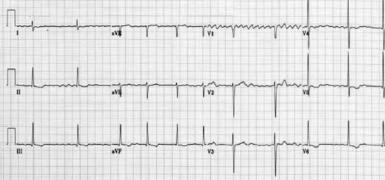
1st degree: Each P-wave is followed by a QRS complex, but the PR interval is prolonged (> 200 ms).

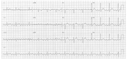
2nd degree: Not all P waves are followed by a QRS complex.

Mobitz Type I: In Mobitz type I (Wenckebach) there is a progressive prolongation of the PR interval (AV conduction) until eventually an atrial impulse is completely blocked. When an atrial impulse is completely blocked there will be a P wave without a QRS complex. This pattern is often referred to as a “dropped beat.”. Here each depolarization results in the prolongation of the refractory period of the atrioventricular node (AVN); hence itis progressive. Eventually, an impulse comes when the AV node is in its absolute refractory period and will not be conducted. This will manifest on the ECG as a P wave that is not followed by a QRS complex. This non-conducted impulse allows time for the AV node to reset, and the cycle continues. When seen in young, fit people with high vagal tone, often nocturnally, it may be benign. However, in the elderly it can be a sign of significant AV node disease and there may be a risk of inducing higher degrees of AV block, particularly with AV nodal blocking agents (e.g. beta-blockers, verapamil, diltiazem, amiodarone).

Mobitz Type II: In Mobitz type II there is a constant PR interval across the rhythm strip both before and after the non-conducted atrial beat. Each P wave is associated with a QRS complex until there is one atrial conduction or P wave that is not followed by a QRS.

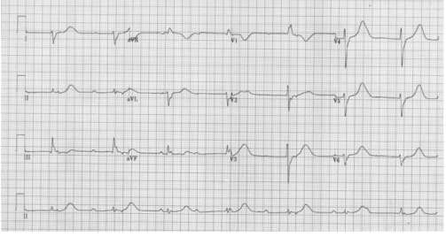
3rd degree : There is complete dissociation of P-waves and QRS complexes due to complete block at the AV node (complete HB). The QRS rate is slower than the P-wave rate as the ventricular complexes are generated by an escape rhythm within the ventricular conduction system, which has a lower intrinsic automaticity rate compared to atrial tissue. If the escape rhythm originates high within the His-Purkinje system, the escape rhythm may consist of narrow QRS complexes (since onward ventricular depolarisation occurs via the normal conduction pathway) and only slightly reduced HR (40–50 bpm). The lower the escape rhythm in the His-Purkinje system, the broader and
slower the QRS complexes and the higher the risk of progressing to asystole

MANAGEMENT

–> For bradycardia, in a symptomatic patient give atropine 500 µg IV, and if the patient remains symptomatic, repeat this dose every 3–5 minutes up to a maximum of 3 g. If there has been a calcium antagonist or beta-blocker overdose consider using glucagon. If the patient is at risk of developing asystole, transvenous pacing may be required. Risk factors for a bradycardia deteriorating to asystole include a recent episode of asystole, Mobitz type II AV block, third-degree (complete) heart block or ventricular standstill of more than three seconds.

–> Complete heart block with narrow complexes may not require immediate pacing, as AV junctional ectopic pacemakers can provide a reasonable and stable cardiac output.

–> Transvenous pacing is the definitive treatment, but if the expertise is not readily available, patients may be managed temporarily with either transcutaneous pacing or an epinephrine infusion in the range of 2–10 µg per minute.

–> If there is no pacing equipment immediately available, fist pacing may be attempted. Apply serial rhythmic blows with a closed fist over the left sternal edge to pace the heart at physiological rate of 50–70 times a minute

INDICATIONS FOR TEMPORARY PACING IN THE PERIOPERATIVE PERIOD

  • Complete heart block
  • Second degree heart block
  • First degree heart block with left bundle branch block
  • Trifascicular block

OTHER INDICATIONS FOR TEMPORARY PACEMAKER

  • Bradycardia unresponsive to atropine
  • Inferior wall MI which can impair perfusion to AV node
  • Anterior wall MI affecting bundle branches in the IVS
  • Overdrive pacing for AV reentry tachycardia and VT
  • Post aortic or VSD or tricuspid surgery
  • Asystole with p wave activity

FIRST DEGREE HEART BLOCKScreen Shot 2019-04-23 at 8.37.53 PM.png

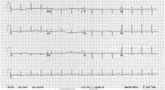
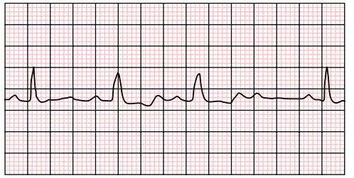
SECOND DEGREE HEART BLOCK

Screen Shot 2019-04-23 at 8.38.46 PM

COMPLETE HEART BLOCK

Screen Shot 2019-04-23 at 8.38.13 PM

Broad Complex Tachycardias

A broad complex tachycardia has a QRS complex greater than 0.12 seconds. They are usually ventricular in origin, but can also be supraventricular with aberrant conduction. Other possible causes for broad complex tachycardias include atrial fibrillation with ventricular pre-excitation, i.e. patients with Wolff–Parkinson–White (WPW) syndrome, or torsades de pointes (polymorphic VT).

Broad complex tachycardia is therefore due to SVT with aberrancy or Ventricular Tachycardia (VT), and differentiating between the two can be challenging. However, there are a few pathognomonic ECG features that diagnose VT

Screen Shot 2019-04-22 at 9.35.57 PM

1.Atrio-ventricular (AV) dissociation. There is a higher ventricular rate than atrial rate (more QRS complexes than P-waves). This can only occur if the ventricular rate is autonomous and no longer under control of the SA node.

2.Capture beats: There is an isolated narrow complex amongst a train of broad complexes. This represents a normally conducted P-wave via the AV node and an intact His-Purkinje system indicating there is no underlying bundle branch block. Therefore, the train of broad complexes are ventricular in origin (i.e. VT).

3.Fusion beats: A normally conducted P-wave may fuse with a simultaneous ventricular beat causing a complex halfway between the appearance of a normal QRS and a broad complex.

4.VT is more likely in patients with a prior history of MI.

5.VT complexes are usually very broad (> 160 ms) due to a very abnormal path taken by the depolarisation wave from the VT focus.

6.The time from R-wave onset to the nadir of the S-wave is prolonged (> 100 ms) in VT, again representing an abnormal activation path through the ventricle.

7.Extreme left axis deviation and positive aVR are more common in VT, as the ventricles are depolarised in the opposite direction to normal conduction.

8.Failure to respond to iv adenosine

9.The absence of typical RBBB or LBBB patterns suggests VT. For example, an RSR pattern in V1 with a taller first R-wave suggests VT (in RBBB the first R-wave is caused by septal depolarisation and is therefore smaller than the second R-wave, which is caused by depolarisation of the RV).

SVT with aberrancy is more likely if previous ECGs demonstrate an accessory pathway or a bundle branch block with identical morphology to the broad complex tachycardia. When in doubt, treat as VT

TORSADES DE POINTES

Is a specific variant of ventricular tachycardia (VT). It has a classic undulating pattern with variation in the size of QRS complex. It is caused by a prolonged QT interval and can precipitate VF and sudden death

QT PROLONGATION: CAUSES

Tricyclic antidepressants, flecainide and quinidine; Hypocalcemia; Acute myocarditis

VENTRICULAR TACHYCARDIA(VT) AND VENTRICULAR FIBRILLATION (VF)

VT is a broad complex tachycardia, defined as a run of at least three consecutive ventricular ectopic beats, at a rate of >120 bpm. Can arise from a single or multiple foci or from a reentry circuit. There may be capture or fusion beats, where a normally conducted beat will join an ectopic beat travelling in the opposite direction

CAUSES: Acute MI, degeneration of other arrhythmias, electrolyte abnormalities etc

VF describes an ECG which is random and chaotic with no identifiable QRS complexes that is incompatible with life and need immediate provision of ACLS with prompt delivery of DC shock. Others: Amiodarone, Lidocaine, beta blockers, Implantable cardioverter defibrillators


MANAGEMENT

For VT treat with amiodarone 300 mg IV followed by 900 mg over 24 hours. If the arrhythmia is known to be supraventricular, treat as a narrow complex tachycardia.

An irregular broad complex tachycardia is most likely to be atrial fibrillation with bundle branch block, and should be treated as narrow complex atrial fibrillation

In a stable patient who is known to have WPW, the use of amiodarone is probably safe. Adenosine, digoxin, verapamil and diltiazem must be avoided, as these drugs block the AV node and will cause a relative increase in pre-excitation

Torsades de pointes is treated by stopping all drugs known to prolong the QT interval and correcting electrolyte abnormalities. Magnesium sulphate (2 g IV over 10 minutes) should also be given. Such patients may require ventricular pacing. If the patient’s condition deteriorates proceed to synchronised electrical cardioversion or, if the patient is pulseless, commence the ALS algorithm

Ventricular bigeminy
Ventricular bigeminy is associated with endotracheal intubation (a sympathoadrenal response). Given time the bigeminy will disappear, but if it does not intravenous
lidocaine (50–100 mg) may be helpful

Narrow Complex Tachycardias

Narrow complex tachyarrhythmias have a QRS duration <0.12 seconds. They arise above the bundle of His.

NARROW COMPLEX TACHYCARDIA

As narrow complex tachycardias involve ventricular activation through the normal His-Purkinje system, they must originate within the atria and are therefore often referred to as supraventricular tachycardia (SVT). There are five common types of SVT. They are: Atrial tachycardia, Atrial fibrillation, Atrial flutter, Atrioventricular nodal
re-entry tachycardia, Atrioventricular re-entry tachycardia. When faced with an ECG of narrow complex tachycardia, (i) we should examine the P-wave and (ii) check the QRS regularity

SINUS ARRHYTHMIA/TACHYCARDIA/BRADYCARDIA (from SA Node)

ATRIAL FIBRILLATION 

There is completely disorganised atrial activity, with P-waves replaced by an irregular baseline due to fibrillation waves, and QRS complexes occur in an irregularly irregular fashion (Please the post on AF)

Screen Shot 2019-04-18 at 9.23.35 PM

ATRIAL FLUTTER 

There is a self-perpetuating wave of atrial depolarisation usually circulating within the right atrium, causing regular, saw-toothed flutter waves at 300 bpm and QRS complexes every second, third, or fourth flutter wave. We can see classical sawtooth flutter waves.Drug control of the ventricular rate is not often successful.

Screen Shot 2019-04-22 at 8.06.10 AM

ATRIAL TACHYCARDIA 

There is an abnormal atrial focus driving the ventricular rate. This rhythm can be difficult to distinguish from sinus tachycardia, but P-wave morphology and axis is usually abnormal. If the atrial focus is close to the AV node, a junctional tachycardia may occur and P-waves may be absent.

In case of Atrial tachycardia with AV block after halting glycoside therapy (and ensuring normokalaemia), lidocaine 1 mg kg−1 IV is the drug
of choice. Alternatively DC cardioversion or atrial
pacing may be effective.

ATRIO VENTRICULAR NODAL REENTRY TACHYCARDIA (AVNRT)

This is the commonest type of paroxysmal supraventricular tachycardia (PSVT). It is often seen in people without any heart disease, and is usually benign. There is a rapid reentry circuit within the AV node resulting in simultaneous atrial and ventricular depolarisation. The P-wave is usually buried within the QRS or ST-segment. There will be fast regular narrow complex tachycardia, and P-waves can be seen buried in the terminal portion of the QRS complex which may easily be mistaken for a second, small R-wave. The very close proximity of the QRS and P-waves implies near simultaneous depolarisation of atria and ventricles.

Screen Shot 2019-04-22 at 8.23.38 AM

ATRIO VENTRICULAR REENTRY TACHYCARDIA (AVRT)

This occurs in patients with WPW, and is usually benign unless there is coexisting structural heart disease. There is an accessory pathway bridging the atria and ventricles allowing antegrade conduction down the AV node (causing a narrow QRS) and retrograde conduction back to the atria via the accessory pathway. Since the depolarisation wave takes time to complete this circuit, the P-wave occurs after the QRS complex and is often buried within the T-wave. AVRT can occur with antegrade conduction to the ventricles via the accessory pathway, but this will result in ventricular depolarisation via an abnormal route and consequently a broad QRS. In sinus rhythm, antegrade conduction via the accessory pathway produces a short PR interval (as the normal delay in the AV node is avoided) and the abnormal activation of the ventricles produces a slurred upstroke
in the QRS called a delta wave. The QRS complex is said to be pre-excited and can be associated with repolarisation abnormalities. There are seven sinus beats followed by a ventricular ectopic beat that conducts to the atria retrogradely through the atrioventricular node and then returns to the ventricles via the accessory pathway. This cycle repeats and triggers a broad complex tachycardia. (Please see post on ‘WPW Syndrome’ also).

Screen Shot 2019-04-22 at 8.44.52 AM

An unstable patient presenting with a regular narrow complex tachycardia should be treated with electrical cardioversion. If this is not immediately available, adenosine should be given as a first-line treatment. A stable patient presenting with a regular narrow complex tachycardia should initially be treated by vagal
manoeuvres such as carotid sinus massage or the Valsalva manoeuvre, as these will terminate up to a quarter of episodes of PSVT. Carotid sinus massage should be avoided
in the elderly, especially if a carotid bruit is present, as it may dislodge an atheromatous plaque and cause a stroke

Management

A stable patient presenting with a regular narrow complex tachycardia should initially be treated by vagal manoeuvres such as carotid sinus massage or the Valsalva manoeuvre, as these will terminate most episodes of PSVT. Carotid sinus massage should be avoided in the elderly, especially if a carotid bruit is present, as it may dislodge an atheromatous plaque and cause a stroke. If the tachycardia persists and is not atrial flutter, 6 mg of adenosine should be given as an IV bolus, followed by a 12 mg bolus if no response. A further 12 mg bolus of adenosine may be given if the tachycardia persists. Vagal manoeuvres or adenosine will terminate almost all AVNRTs or AVRTs within seconds, and therefore failure to convert suggests an atrial tachycardia such as atrial flutter. If adenosine is contraindicated, or fails to terminate a narrow complex tachycardia, without first demonstrating it as atrial flutter, give a calcium-channel blocker, e.g. verapamil 2.5–5 mg IV over two minutes. Atrial flutter should be treated by rate control with a beta-blocker.

An irregular narrow complex tachycardia is most likely to be atrial fibrillation (AF) with an uncontrolled ventricular response, but may also be atrial flutter with variable block. If the patient is unstable, synchronised electrical cardioversion should be used to treat the arrhythmia

ATRIAL FIBRILLATION (AF) AND THE ANESTHESIOLOGIST

Atrial fibrillation(AF) is a supra-ventricular arrhythmia characterized by the complete absence of co-ordinated atrial contractions. There will not be any discernable p-waves.
The ventricular response rate depends on the conduction of the AV node.

WHAT IS THE DIFFERENCE BETWEEN ATRIAL FIBRILLATION AND ATRIAL FLUTTER

Flutter is a more organised and regular form of atrial activity and classically with an atrial rate of 300 bpm. ‘Saw toothed’ flutter waves are present on the ECG. The ventricular response depends on conduction through the AV node. The classic ECG has 2:1 block, hence a ventricular rate of 150 bpm

CAUSES OF AF IN THE PERIOPERATIVE SETTING

Electrolyte abnormalities especially low potassium or magnesium
Withdrawal of beta blockers
Following cardiac surgery.
ASD or mitral valve disease
Ischaemic heart disease
Thyrotoxicosis
Excess caffeine or alcohol (acute or chronic)
Pulmonary embolism
Pneumonia
Pericarditis

In the context of major vascular surgery, systemic inflammation,hypovolemia and a heightened adrenergic state are likely to play a major role.

WHAT IS LONE AF?

‘Lone AF’ is AF in the absence of any demonstrable medical cause, but this is not usually diagnosed in the peri-operative period. So beta blockers will be efficacious in this setting.

WHAT ARE THE PROBLEMS AF CAN POSE?

Loss of the atrial ‘kick’ as it contracts and empties into the LV can reduce the CO by 10%–20% with a normal ventricle (reduced by 40%–50% in those with a ‘stiff’ ventricle as in
diastolic dysfunction, aortic stenosis etc). The disorganised contractions of the atria cause stasis of blood and the risk of thromboembolism. There is a 3%–7% annual risk of
thromboembolic CVA

AF- EVALUATION

*History *Assessment of volume status and electrolytes *ECG: This will also help to exclude acute ischaemia. *The pulse will be irregularly irregular. *No ‘a wave’ in the jugular venous pulsation as this is caused by sinus atrial contraction. *Chaotic atrial activity can be seen on echocardiography.

READ THIS ECG

Screen Shot 2019-04-18 at 9.23.35 PM.png

MNEMONIC ‘RIAS QRST’ (Rate & Rhythm, Intervals, Axis, QRS & ST segment T wave)

The rate is 78 bpm; the rhythm is irregularly irregular. There are flutter waves seen in the V1 rhythm strip. The axis is normal (There is borderline LVH by voltage criteria). There are no Q waves and the QRS width is normal. There is evidence of infero-lateral ischaemia shown by the inverted and biphasic T waves in this territory (II, III, aVF and V3−V6).

MANAGEMENT OF AF

Assess for cardiovascular compromise and resuscitate simultaneously if needed. Oxygen should be administered, continuous ECG monitoring instituted and IV access secured.

If the patient is unstable, synchronised electrical cardioversion should be used to treat the arrhythmia.

In a stable patient, treatment options include: Rate control by drug therapy. Drugs used to control the heart rate include beta-blockers, digoxin, magnesium, the non-dihydropyridine calcium channel blockers (verapamil or diltiazem) or a combination of these.

Rhythm control by amiodarone to encourage cardioversion: Amiodarone is given as a 300 mg IV bolus, followed by 900 mg IV over 24 hours.

Rhythm control by electrical cardioversion: This is more likely to restore sinus rhythm than chemical cardioversion.

Treatment to prevent complications. Patients who are in AF are at risk of atrial thrombus formation and should be anticoagulated

Patients who have pre excitation syndromes with an accessory conduction pathway between the atria and ventricles (such as in the Wolff–Parkinson–White syndrome) should not be given AV node blocking drugs if they develop an SVT. This will promote the atrial impulses to travel directly to the ventricle at up to 300 bpm via the accessory pathway. The drugs of choice are amiodarone, flecainide or procainamide.

BEFORE PROCEEDING WITH DC CARDIOVERSION FOR AF, WHAT ALL THINGS SHOULD BE CONSIDERED?

  1. Cardioversion should only be attempted without anticoagulation if the duration of the AF is less than 48 hours. If the duration is unknown or longer than this, 3–4 weeks of anticoagulation (INR 2–3) is required to reduce the incidence of clot embolisation. If there is a contra-indication to anticoagulation, or if the cardioversion is deemed necessary more urgently, then an echocardiogram is needed to exclude thrombus in the atrium and atrial appendage
  2. When did the AF start (history of palpitations or recording on monitor): is it acute or chronic?
  3. What is the likelihood of an atrial thrombus which could be embolised by
    cardioversion?
  4. What is the ventricular rate now? – may need pacing after cardioversion if
    the rate is below 60 bpm
  5. Has there been an ischaemic episode?

ANESTHESIA FOR CARDIOVERSION

This should be done in a critical care or operating room area with the usual preparation, equipment and assistance needed for any routine anaesthetic. Someone independent should be present to perform the defibrillation, preferably with a hands-free device. Elective cardioversion has been done under conscious sedation without any adverse effects, but the usual technique is to use a sleep dose of propofol following pre oxygenation. One can use a facemask or maintain the airway with an LMA. If there is any serious doubt about cardiovascular performance or reserve, an arterial line should be given consideration, but this is a short procedure and the cardiac output should improve with the restoration of sinus rhythm. If the patient has a pacemaker in situ or an implantable cardiac defibrillator, we should place the paddles as far away as possible from the device and preferably in the anterior–posterior position.

IF THE PATIENT DOES NOT GET CARDIOVERTED, WHAT SHOULD YOU DO?

Try a period of 4–6 weeks of medical therapy and anticoagulation. If the patient is still in AF, then a further trial of DCC is reasonable. If a second DCC is unsuccessful, then rate control is the next step to improve symptoms and reduce ventricular failure.


Wolff–Parkinson–White syndrome and the Anesthesiologist

WHAT ARE THE CAUSES OF PALPITATION

Exercise, Anxiety, Caffeine, alcohol, drugs: thyroxine, cocaine, beta 2 agonists, MI, arrthymias, hyperthyroidism, hypoglycaemia, phaeochromocytoma

MECHANISMS OF ARRHYTHMIAS

Reentry circuits, Enhanced automaticity, Triggered activity

EVALUATION AND MANAGEMENT

History and examination, ECG: 12-lead, 24-hour, ambulatory, cardiac electrophysiological study, blood investigations to rule out endocrine causes

ABC, oxygen, etc., Check electrolytes (including Mg2+), Carotid sinus massage, Adenosine – caution in asthma and if taking dipyridamole prolongs half-life

WHAT HAPPENS IN WPW SYNDROME?

Presence of faster accessory pathway (bundle of Kent) between atrium  and ventricle (accessory AV pathway) which conducts impulses faster than the normal AV node. Electrical signals traveling down this abnormal pathway may stimulate the ventricles to contract prematurely, resulting in a unique type of supraventricular tachycardia. The ECG may show sinus rhythm, normal axis, short PR interval and the presence of delta waves

DELTA WAVES

The accessory atrio-ventricular pathway conducts the atrial impulse to the ventricles much faster than the A–V node. This results in the start of ventricular depolarisation sooner than normal, hence the short P–R interval. That initial ventricular depolarisation
takes place in normal ventricular tissue (i.e. not specialised conducting tissue). The initial rate of depolarisation is therefore slower, hence the slurred, delta wave. When the rest of the impulse finally arrives the A–V node, the bundle of His and Purkinje carries out the ventricular depolarization as normal; hence, the rest of the QRS looks normal

IMPLICATIONS FOR ANESTHESIA

1.There is a tendency to paroxysmal supraventricular tachycardia in the perioperative period and there may be associated congenital cardiac abnormality.

2.Anaesthetic drugs tend to change the physiology of AV conduction.

3.If the patient is asymptomatic, then risk of perioperative arrhythmias is much less.

4.We should avoid light planes of anaesthesia and drugs that can precipitate tachycardia (like atropine, glycopyrrolate, ketamine) resulting in paroxysmal supraventricular tachycardia or atrial fibrillation

5.There are references showing disappearance of delta waves after propofol administration, making it the drug of choice for induction. For maintenance, Isoflurane and sevoflurane are preferred as they dont have effect on AV node conduction. Short acting nondepolarizing muscle relaxant would be an acceptable choice as reversal of neuromuscular blockade using neostigmine and glycopyrrolate is not required.

6. Regional anaesthesia has significant advantage over general anaesthesia as multidrug
administration, laryngoscopic stimulation, intubation, and light planes leading to sympathetic stimulation are avoided.

WHAT ARE THE COMMON ARRHYTHMIAS IN WPW?

Atrial fibrillation (AF): Patients with WPW who develop atrial fibrillation are at risk of very rapid ventricular responses as the accessory pathway does not provide any ‘protective delay’ like the A-V node. This may result in heart failure or may even deteriorate into ventricular fibrillation. In AF, most conducted impulses reach the ventricles via the accessory pathway, so delta waves are seen on the ECG.
Re-entrant tachycardia: A re-entry circuit is set up. After transmitting an atrial impulse, the A–V node usually recovers before the accessory pathway. If an atrial ectopic occurs at the right time, it will transmit through the A–V node while the accessory pathway is still refractory. By the time it has done this, the accessory pathway may have recovered and the impulse will then pass through it back into the atria. As the impulses are all reaching the ventricles via the A–V node and not the accessory pathway, there are no delta waves on the ECG

INTRAOPERATIVE ARRHYTHMIAS: MANAGEMENT

1. A,B,C and Treat possible triggers of rhythm disturbance such as hypoxia, hypercarbia, acidosis, electrolyte disturbance or any cause of sympathetic stimulation.

2.Assess the degree of cardiovascular compromise. If there is significant compromise, synchronised DC cardioversion starting at 25–50 J would be the treatment of choice. If the blood pressure was stable, then the management would depend on the rhythm.

3. Pharmacological therapy:

(a) For re-entrant tachycardia, adenosine would be the first choice. Class 1a drugs such as procainamide (5–10 mg/kg) and disopyramide prolong the refractory period, decrease conduction in the accessory pathway (by blocking fast sodium channel) and may terminate both re-entrant tachycardia and AF. More conventional drugs such as amiodarone, sotalol and other beta-blockers such as esmolol may also be useful.

(b)AF: The treatment principle is to prolong the anterograde refractory period of the accessory pathway relative to the AV node. This slows the rate of impulse transmission through the accessory pathway and, thus, the ventricular rate. This is in direct contradiction to the goal of treatment of non-WPW atrial fibrillation, which is to slow the refractory period of the AV node

DRUGS THAT SHOULD BE AVOIDED

Verapamil and digoxin are contra-indicated as they both preferentially block A–V conduction thereby increasing conduction through the accessory pathway. Although verapamil could, in theory, be used to terminate a re-entrant tachycardia, its use is not advisable, because these patients may then revert to AF or flutter. A further hazard with verapamil is that a tachyarrhythmia that looks like re-entrant tachycardia may actually be VT. Adenosine would preferentially block the A–V node and therefore should not be used in AF.