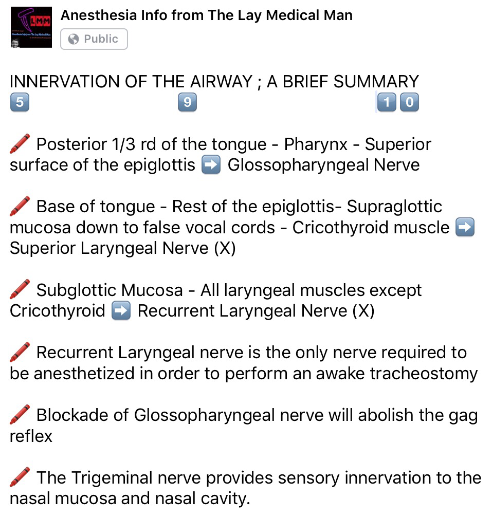
VIVA SCENE: RADIAL NERVE COURSE AND CAUSES OF INJURY

COURSE:

  • The radial nerve (C5–8, T1) transmits fibres from all the roots of the brachial plexus.
  • At its origin it lies behind the third part of the axillary artery; it then passes between the long and medial heads of triceps into the posterior compartment of the arm, accompanied by the profunda branches of the brachial vessels.
  • It descends first along the spiral groove of humerus, and then between muscle planes
  • A hand’s breadth above the elbow, the nerve reaches the lateral margin of the humerus, and enters once more into the anterior compartment of the arm, where it lies between brachialis and brachioradialis. At this point, the nerve is susceptible to compression injury, in particular from an arterial tourniquet placed too low around the arm.
  • It ends in front of the lateral epicondyle of the humerus by dividing into two terminal branches, the superficial radial nerve and the posterior interosseous nerve.

BRANCHES:

The muscular branches:

  • Medial group (arising in the axilla): Supplies: a long head of triceps b medial head of triceps.
  • Posterior group (arising in the spiral groove) : a medial head of triceps b lateral head of triceps c anconeus.
  • Lateral group: a brachialis (together with musculocutaneous nerve) b brachioradialis c extensor carpi radialis longus

The cutaneous branches:

  • The posterior cutaneous nerve of the arm, which arises in the axilla and supplies the skin over the proximal one-third of the posterior aspect of the arm.
  • The posterior cutaneous nerve of the forearm, which arises in the spiral groove,  supplies the skin over the posterolateral aspect of the forearm.
  • The lower lateral cutaneous nerve of the arm, supplies an area of skin over the lateral aspect of the arm just above the elbow.

The posterior interosseous nerve (terminal branch), passes into the posterior compartment of the forearm. It supplies supinator, many extensors and abductor pollicis longus. It also supplies the wrist joint.

The superficial radial nerve (terminal branch) is entirely sensory. It divides into dorsal digital nerves and supply the dorsal aspect of the hand upto the radial half of the ring finger.

CAUSES OF RADIAL NERVE INJURY:

  • Saturday Night Palsy: Caused by prolonged compression of the nerve at the spiral groove.
  • Mechanical compression of the radial nerve in the spiral groove can also occur as a result of the continuous use of crutches or prolonged kneeling in a “shooting” position
  • As a delayed complication of a chronic intramuscular injection leading to muscle fibrosis
  • From prolonged inflation of an automatic blood pressure cuff especially when placed over the distal third around the arm, in a lean patient. Here, the radial nerve lies in direct contact with the humerus and there are no muscle fibers to act as a cushion between the nerve and the periosteum of the bone
  • Humeral shaft fracture

LUMBAR PLEXUS BLOCK

Anatomy of lumbar plexus?

The lumbar plexus is formed by the anterior primary rami of L1 to L4 (Sometimes T12 also may contribute)
L1 forms the iliohypogastric and ilioinguinal nerves
It also gives a contribution to the formation of the genitofemoral nerve
L2 forms the lateral cutaneous nerve of thigh, along with L3
The obturator and femoral nerves are formed by contributions from L2,L3,L4

What are the indications?

Surgeries involving hip/thigh/upper leg/trauma
In conjunction with sciatic nerve or sacral plexus block
Cancer pain arising from hip or upper femur
Sympathetic block also helps in ischemic pain and CRPS

Which approach will you use?

I will use the 3 in 1 approach that aims to block the femoral, obturator and lateral cutaneous nerves with a low anterior approach. The patient is in the supine position.I will use a nerve stimulator. Using a 50 mm insulated needle, I will puncture the skin at a point 1 cm lateral to the femoral pulse and 2 cm below the inguinal ligament, at 45 degrees to skin and directed proximally and parallel to the femoral artery. The endpoint is a quadriceps twitch, which occurs at a depth of 30-50 mm. I will press distal to the injection to enhance the spread of the drug proximally. I will block the lateral cutaneous nerve separately by injecting 10 ml of local anesthetic at a point 2 cm inferior and medial to the ASIS. Other approaches are psoas compartment block and fascia iliacus block

How does the lumbar sympathetic block differ from the lumbar plexus block?

The lumbar sympathetic nerves lie anterolateral to the vertebral body, whereas the somatic nerves lie posterior to the psoas muscle and fascia. The sympathetic chains have anteriorly, aorta on the left and IVC on the right. The aim is to deposit local anesthetic around the nerves from L2 to L4 either with a single injection at L3 or 3 separate injections at L2, L3 and L4. It is used in lower limb ischemia, CRPS, phantom limb, urogenital pain etc

How to do the lumbar sympathetic block?

Patient should be positioned lateral with the side to be blocked up. A point is marked 8 cm from the midpoint of the spinous process of the desired vertebra. Its done under image guidance. A 12 cm 22 G needle is inserted at 45 degree angle directed medially towards the vertebral body which lies at a depth of around 8 cm; if the needle hits the bone at 4-5 cm depth it is likely to be the transverse process and should be redirected cranially or caudally to pass over it. Once the needle hits the vertebral body, it should be redirected slightly antero-laterally till we feels the pop-off of passing the psoas fascia. Then the local anesthetic mixed with radiographic contrast is injected which should form a band around the desired vertebral bodies. If the contrast disappears very quickly, it may be in a vessel. Otherwise, it can go into the psoas muscle or retroperitoneal tissue. Also we should take lateral and anteroposterior images to confirm the correct position.

Complications of lumbar sympathetic block?

Local anesthetic toxicity due to injection into aorta or IVC
Profound motor block or permanent paralysis due to intrathecal injection
Profound hypotension: good iv access and access for resuscitation equipments are a must
Post sympathectomy pain
Ureteric injury, ejaculatory failure

Screen Shot 2019-03-18 at 3.15.22 PMScreen Shot 2019-03-18 at 11.01.44 AM

VOCAL CORD PALSIES

Under normal circumstances, the vocal cords meet in the midline during phonation. On inspiration, they move away from each other. They return toward the midline on expiration, leaving a small opening between them. When laryngeal spasm occurs, both true and false vocal cords lie tightly in the midline opposite each other.

The recurrent laryngeal nerve (RLN) carries both abductor and adductor fibers to the vocal cords.

Selmon’s law: The abductor fibers are more vulnerable, and moderate trauma causes a pure abductor paralysis. Severe trauma causes both abductor and adductor fibers to be affected. N.B.:- Pure adductor paralysis does not occur as a clinical entity.

Scenario 1- PURE UNILATERAL ABDUCTOR PALSY: As adduction is still possible on the affected side, the opposite cord come and meet in the midline on phonation. However, only the normal cord abducts during inspiration.

Scenario 2- COMPLETE UNILATERAL PALSY OF THE RLN: Both abductors and adductors are affected. On phonation, the unaffected cord crosses the midline to meet its paralyzed counterpart, appearing to lie in front of the affected cord. On inspiration, the unaffected cord moves to full abduction.

Scenario 3- BILATERAL INCOMPLETE ABDUCTOR PALSY: When there is incomplete bilateral damage to the recurrent laryngeal nerve, the adductor fibers draw the cords toward each other, and the glottic opening is reduced to a slit, resulting in severe respiratory distress.

Scenario 4- COMPLETE BILATERAL PALSY OF THE RLN: With a complete palsy, each vocal cord lies midway between abduction and adduction, and a reasonable glottic opening exists.

Thus, bilateral incomplete palsy is more dangerous than the complete variety.

Scenario 5- DAMAGE TO SUPERIOR LARYNGEAL NERVE/S: Damage to the external branch of the superior laryngeal nerve or to the superior laryngeal nerve trunk causes paralysis of the cricothyroid muscle (the tuning fork of the larynx), resulting in hoarseness that improves with time because of increased compensatory action of the opposite muscle. The glottic chink appears oblique during phonation. The aryepiglottic fold on the affected side appears shortened, and the one on the normal side is lengthened. The cords may appear wavy. The symptoms include frequent throat clearing and difficulty in raising the vocal pitch.

Scenario 6- TOTAL BILATERAL PARALYSIS OF VAGUS NERVES: This affects the recurrent laryngeal nerves and the superior laryngeal nerves. In this condition, the cords assume the abducted, cadaveric position. The vocal cords are relaxed and appear wavy. A similar picture may be seen after the use of muscle relaxants.

Screen Shot 2018-06-26 at 11.01.24 PM

N.B:- Topical anesthesia of the larynx may affect the fibers of the external branch of the superior laryngeal nerve and paralyze the cricothyroid muscle, signified by a “gruff” voice. Similarly, a superior laryngeal nerve block may affect the cricothyroid muscle in the same manner as surgical trauma does.

Reference: Benumof and Hagberg’s Airway Management, Third edition

SYMPATHETIC GANGLIA & CHAIN

PREGANGLIONIC NEURONS of the sympathetic system synapse with the POSTGANGLIONIC NEURONS in the SYMPATHETIC GANGLIA. These ganglia together will form sympathetic chain. The sympathetic chains extend down the length of the vertebral column and are divided into four parts:
CERVICAL SYMPATHETIC GANGLIA

🔸Consist of three ganglia: Superior, Middle and Inferior

🔸Superior sends postganglionic fibres to form the internal carotid plexus

🔸Inferior or Stellate ganglion is fused with the first thoracic ganglia

THORACIC SYMPATHETIC GANGLIA

🔸T1-T5 ganglia supply the aortic, cardiac and pulmonary plexus

🔸The last 7 thoracic ganglia form the greater and lesser splanchnic nerves

🔸The lowest splanchnic nerve is formed from the last thoracic ganglia and supplies the renal plexus

LUMBAR PREVERTEBRAL SYMPATHETIC GANGLIA 

🔸Supplies the coeliac plexus

SACRAL SYMPATHETIC GANGLIA (PELVIC)

🔸Contribute to hypogastric and pelvic plexus

THE PARASYMPATHETIC NERVOUS SYSTEM: POINTS RELEVANT FOR THE ANESTHESIOLOGIST

🎳The anterior hypothalamus controls the parasympathetic nervous system

🎳The posterior and lateral hypothalamic areas increase blood pressure and heart rate, whereas the preoptic area decreases blood pressure and heart rate.

🎳The parasympathetic nervous system arises from neurons in the brainstem and spinal cord sacral segments (S 2 –S 4 ). As the parasympathetic ganglia are located near or within their effector organs, the parasympathetic postganglionic fibres are short, and they all release acetylcholine. The distribution of parasympathetic outflow is restricted so that parasympathetic effects are more localized than sympathetic effects.

🎳Cranial nerves 3, 7, 9 and 10 have parasympathetic components (3 – pupil and ciliary body constriction; 7 – tearing and salivation; 9 -salivation: 10 – the vagus and its ramifications).. Preganglionic fibres of the third cranial nerve arise from the oculomotor nucleus and pass through the orbit to the ciliary ganglion. Postganglionic fibres from the ciliary ganglion supply the ciliary muscle and sphincter of the iris and constrict the pupils.
🎳Preganglionic fibres from the superior salivary nucleus of the seventh nerve form the chorda tympani and reach the submaxillary ganglion via the lingual nerve. Postganglionic fibres supply the submaxillary and sublingual salivary glands and cause salivary secretion. #TheLayMedicalMan
🎳Preganglionic fibres arising from the inferior salivary nucleus of the ninth nerve form the lesser superficial petrosal nerve and reach the otic ganglion. The postganglionic fibres are distributed to the parotid gland via the auriculotemporal nerve and also cause salivary secretion.
🎳The vagus nerve is the major part of the cranial parasympathetic outflow. The preganglionic fibres arise from the dorsal nucleus of the vagus in the medulla and terminate in the ganglia of plexuses or in the walls of visceral organs. Postganglionic fibres supply the heart and decrease cardiac excitability, contractility, conductivity and rate. Postganglionic fibres from the pulmonary plexus contract the circular muscles of the bronchi, producing bronchoconstriction. Vagal branches to the gastric plexus give rise to postganglionic fibres to the stomach, liver, pancreas and spleen. Stimulation of the vagus causes increased gastric motility and secretions, with relaxation of the pyloric sphincter. The intestinal branches of the vagus supply the small and large intestines down to the transverse colon and it’s stimulation increases peristalsis and relaxes the ileocolic sphincter. #TheLayMedicalMan
🎳 The sacral outflow of the parasympathetic system arises from the second, third and fourth sacral segments of the spinal cord, and fibres enter the hypogastric plexus to innervate the descending colon, rectum, bladder and uterus. It’s stimulation contracts the muscular wall of the rectum, relaxes the internal sphincter of the anus and contracts the detrusor muscle of the bladder wall. 
Ref: Principles of Physiology for the Anaesthetist , 3/e
#physiology , #anaesthesia

THE SYMPATHETIC NERVOUS SYSTEM: POINTS RELEVANT FOR THE ANESTHESIOLOGIST

🔸Posterior hypothalamus is the principal site of sympathetic nervous outflow.
🔸The sympathetic system has short preganglionic fibres and long postganglionic fibres
🔸The sympathetic nerves originate from columns of preganglionic neurons in the grey matter of the lateral horn of the spinal cord from the first thoracic segment down to the second or third lumbar segment.
🔸’BASIC CIRCUIT’ : PREGANGLIONIC NEURONS in the grey matter of the lateral horn of the spinal cord (from T1-L3 segment) –> they leave the spinal cord through the VENTRAL roots with the spinal nerves and then leave the spinal nerves as WHITE RAMI COMMUNICANTES (myelinated B fibres)–> they then synapse with the POSTGANGLIONIC NEURONS in the GANGLIA OF THE SYMPATHETIC CHAIN –> These ganglia together will form sympathetic chains –> The POSTGANGLIONIC NEURONS leave the ganglia as GREY RAMI COMMUNICANTES (unmyelinated C fibres) and join the spinal nerves or visceral nerves to innervate the target organ.

🔸In general, the preganglionic fibres secrete acetyl choline as neurotransmitter, while the post ganglionic ones secrete norepinephrine. The postganglionic sympathetic nerves that innervate the blood vessels of muscles, sweat glands and the hair follicles in the skin release acetylcholine instead of norepinephrine (noradrenaline).
➡️ The sympathetic chains extend down the length of the vertebral column and are divided into four parts:
🔸A cervical part consisting of superior, middle and inferior galglia, supplying the head, neck and thorax. The superior cervical ganglion sends postganglionic fibres to form the internal carotid plexus. The inferior cervical or stellate ganglion is fused with the first thoracic ganglion.
🔸Branches of the thoracic part, from T1 – T5 supply the aortic, cardiac and pulmonary plexuses. The greater and lesser splanchnic nerves are formed from the lower seven thoracic ganglia. The lowest splanchnic nerve arises from the last thoracic ganglion and supplies the renal plexus.
🔸The coeliac plexus is derived from the lumbar sympathetic ganglia
🔸The sacral ganglia contribute to the hypogastric and pelvic plexus #TheLayMedicalMan
SYMPATHETIC SYSTEM UNIQUE EFFECTS:
DILATATION / RELAXATION

✔️Pupils

✔️Bronchial smooth muscle

✔️Gut, Urinary bladder (sphinctors constrict)

✔️Skeletal muscle arterioles (Post ganglionic transmitter: ACh)

CONSTRICTION / SECRETION

✔️Levator Plapabrae Superioris

✔️Piloerection hair follicle (Post ganglionic transmitter: ACh)

✔️Muscles of seminal vesicles(ejaculation)

✔️Sweat glands (sweating) (Post ganglionic transmitter: ACh)
#physiology , #anaesthesia

BRIEF FUNCTIONAL ANATOMY OF THE BRAIN

✔️Precentral gyrus contains the primary motor cortex
✔️Premotor cortex lies immediately anterior to Primary motor cortex ( Brodmann’s area 6 on the lateral surface of the frontal lobe.)
✔️Premotor cortex is active in response to EXTERNAL visual or somatic sensory cues (e.g. reaching for an object in full view, or identifying an object by touch alone). Also has role in bilateral postural fixation (e.g. stabilization of the hips during walking).
✔️The supplementary motor area occupies a neighbouring part of Brodmann’s area 6 on the medial surface of the frontal lobe.
✔️The supplementary motor area seems to respond to INTERNAL cues, especially intentions to make voluntary movements (even if the movement is not carried out).
✔️Lesions of the supplementary motor cortex are associated with contralateral inability to initiate movements (akinesia).
✔️The inferior frontal gyrus of the dominant hemisphere (usually the left) contains the (Broca’s) motor speech area (Brodmann’s areas 44 and 45)
✔️The somatic sensory cortex, occupies the entire postcentral gyrus (Brodmann’s areas 3, 1 and 2, rostral to caudal).
✔️ Caudal to the somatosensory cortex is the sensory association cortex, which is divided into a superior and an inferior parietal lobule by an intraparietal sulcus.
✔️ The superior lobule is thought to be responsible for conscious awareness of the contralateral half of the body. Lesions of this part of the cortex may result in neglect of the contralateral side of the body.
✔️ The inferior lobule in the dominant (usually left) hemisphere is associated with language functions.
✔️ The cortex adjacent to the parieto-occipital sulcus and the calcarine sulcus, which lies on the medial surface of occipital lobe, constitutes the primary visual cortex (Brodmann’s area 17).
✔️ The superior surface of the temporal lobe contains the primary auditory cortex (Brodmann’s areas 41 and 42), responsible for the conscious perception of sound. Unilateral lesions of the primary auditory cortex will therefore cause partial deafness in both ears.
✔️ The auditory association cortex, known as Wernicke’s area corresponds to Brodmann’s area 22 in the dominant hemisphere. It allows for understanding of the spoken word, and connects with other language areas of the brain.
✔️ The temporal lobe curls inward to form the hippocampus which lies in the floor of the inferior horn of the lateral ventricle, and forms part of the limbic system. Its functions relate to short-term memory and the emotional aspects of behaviour.
✔️ Lying close to the anterior end of the hippocampus is the amygdala. Its a mass of subcortical grey matter that also forms part of the limbic system. It is associated with the conscious appreciation of smells.
✔️ The insula is one of the cortical centres for pain, and is also involved in involuntary activities such as the control of viscera by the autonomic nervous system.
✔️ The basal ganglia includes
• striatum (caudate nucleus, putamen of the lentiform nucleus and the nucleus accumbens) 

• pallidum (globus pallidus of the lentiform nucleus) 

• subthalamic nucleus 

• compact part of the substantia nigra 
✔️ Four basic circuits are known to occur from the cerebral cortex, through the basal ganglia and back to the cortex, by a direct or indirect route: 

-a motor loop is concerned with learned movements;

-a cognitive loop with motor intentions; 

-a limbic loop with emotional aspects of movement and 

-an oculomotor loop with voluntary saccades.
✔️ Limbic system is composed of the the parahippocampal and cingulate gyri, the septal area, the hippocampal formation and the amygdala. Declarative memory (i.e. of new facts and events) may occur as modifications of synapses within the hippocampus.
✔️ Diencephalon comprises, from superior to inferior, the epithalamus, thalamus, subthalamus and hypothalamus.
✔️ The thalamus is the largest component of the diencephalon and is composed of numerous nuclei.
✔️ The epithalamus consists principally of the pineal gland that secretes melatonin and is involved with circadian rhythm and regulation of the onset of puberty and the habenular nuclei.
✔️ The subthalamus contains the subthalamic nucleus which connects to the globus pallidus and substantia nigra and is involved with the control of movement.
✔️ The hypothalamus has important connections with the limbic system, a controlling influence on autonomic nervous system activity and a role in neuroendocrine function.
✔️ The cerebellum coordinates movement by maintenance of equilibrium, posture and muscle tone at an unconscious level. Also concerned with muscular coordination, including trajectory, speed and force of movements.
✔️ The tectum (roof) of the midbrain is formed by four colliculi –the paired superior colliculi are associated with the visual system, and the inferior colliculi with the auditory system.


#FunctionalAnatomy, #NeuroAnatomy, #NeuroAnesthesia, #Neurosurgery ,#brain
Reference: The brain: functional divisions, Leo Donnelly, Neurosurgery, Neuroradiology & Neurocritical Care in Anaesthesia