VIVA SCENE: SUBCUTANEOUS EMPHYSEMA – RADIOLOGY

Subcutaneous emphysema, refers to gas in the subcutaneous tissues

Clinically it is felt as crepitus

In the trauma situation, its presence indicates possible serious injuries that do require urgent management.

RADIOGRAPHY

CHEST X-RAY

There are often striated lucencies in the soft tissues that may outline muscle fibers. It can outline the pectoralis major, giving rise to the ginkgo leaf sign. Often there are displaced rib fractures indicating a cause of the gas.
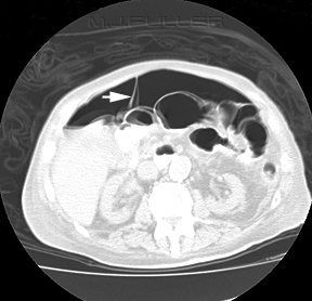
CT

Subcutaneous emphysema is visible on CT scans, with pockets of gas seen as extremely dark low attenuation areas in the subcutaneous space.

USG

Well defined comet tail artefacts can be seen

VIVA SCENE: CT ABDOMEN SHOWING FREE AIR

  • The diagnosis of GI tract perforation is based on the direct CT findings, such as discontinuity of the bowel wall and the presence of extraluminal air, and on the indirect CT findings, such as bowel wall thickening, abnormal bowel wall enhancement, abscess and an inflammatory mass adjacent to the bowel
  • The air may not be contained within any visible bowel wall.
  • Direct visualization of the discontinuity of the bowel wall can specify the presence and site of GI tract perforation, which is marked by a low-attenuating cleft that usually runs perpendicular to the bowel wall on CT
  • The falciform ligament may become outlined with air in a supine patient with free abdominal gas.
  • Screen Shot 2019-09-08 at 10.20.47 pm
  • Screen Shot 2019-09-08 at 10.33.45 pm

Paravertebral Blocks


  • A paravertebral block is essentially a unilateral block of the spinal nerve, including the dorsal and ventral rami, as well as the sympathetic chain ganglion. These blocks can be performed at any vertebral level. However, they are most commonly performed at the thoracic level because of anatomic considerations.
  • They provide analgesia for ✔️Unilateral thoracic pain ✔️Rib fracture ✔️Refractory angina✔️Hyperhydrosis etc
  • Usually a single level injection may cover less than four dermatomes
  • Can be given under USG guidance or using a landmark technique
  • Point to be marked at a point 25 mm lateral to the spinous process of the level to be blocked
  • After local anesthetic infiltration an 18 G epidural catheter is inserted to a depth, not greater than 35 mm till transverse process are hit (they are fairly superficial) and then the needle should be walked off the transverse process caudally, until it is 10mm deeper than the depth at which bone was initially contacted. (cranial walking of the needle increases the chance of pneumothorax)
  • A loss of resistance to injection when the costotransverse ligament is passed is a clue to achieving of correct needle position,; but this is not as marked as the loss of resistance achieved during epidural insertion.
  • If using a peripheral nerve stimulator, contraction of intercostal muscle or transverse abdominis may be elicited
  • 3-5 mL of ropivacaine or levobupivacaine can be used per level. Addition of Clonidine may prolong the blockade