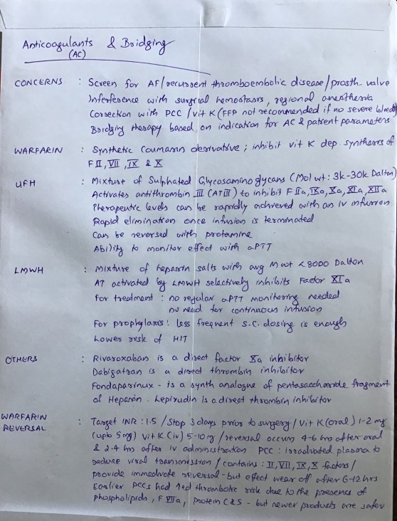
A FEW CLUES IN INTERPRETING AN ISOLATED PROLONGATION OF ACTIVATED PARTIAL THROMBOPLASTIN TIME (aPTT)

aPTT tests the intrinsic and common pathways of coagulation

Though it is included commonly as a part of coagulation profile assessment, it’s primary uses are to detect coagulation factor deficiency and titration of heparin therapy

An isolated elevation of aPTT may indicate

deficiency of Factor VIII or IX or XI or XII

acquired clotting factor inhibitors

presence of Lupus anticoagulant

N.B.:- Factor VIII deficiency is Haemophilia A, Factor IX deficiency is Haemophilia B and Factor XI deficiency is Haemophilia C

If factor levels are >30% of normal, aPTT may remain normal, for e.g. in mild von Willebrand disease [raised aPTT + prolonged Bleeding Time (BT)], in mild hemophilia etc

Reference: Martlew V. Peri-operative management of patients with coagulation disorders. Br J Anaesth. 2000; 85(3): 446–455.

HOMOCYSTINURIA : Anesthesia IMPLICATIONS

There is increased levels of homocystine and methionine in blood and urine due to the deficiency of Cystathionine B synthetase which catalyses the conversion of homocystine and serine into cystathionine

Raised cystine levels reduce the resistance of endothelium against thrombosis, reduces the activity of the vasodilator nitric oxide (NO) and increase platelet aggregation. So there is high incidence of thromboembolism. We have to ensure good hydration, good cardiac output,early mobilisation and should provide mechanical +/- pharmacological thromboprophylaxis. Many patients will be on anticoagulation. If untreated 50% of patients will have thromboembolic complications and the mortality is about 20% before the age of 30 years. So both modification of the dosing of anticoagulants ( especially if regional anesthesia is planned) if patient is receiving them and providing prophylaxis against DVT are important elements of perioperative care. The incidence of thrombotic complications are more in pregnant patients.

Blood viscosity and platelet adhesiveness can be reduced by dextran, and the prior administration of pyridoxine

Reduced cystine results in weak collagen and fragmentation of elastic tissue of large arteries. There is high incidence of vascular diseases like Cerebrovascular diseases, Coronary Artery Disease, Peripheral Vascular Diseases

Patients may have increased insulin levels resulting in hypoglycemia. Dextrose infusion will prevent hypoglycaemia.

Acute psychiatritc symptoms, delirium etc have been reported and the altered availability of homocysteine, methionine and cystiene which are having glutamate agonist properties, has been postulated as a factor which promotes this.

Regional anaesthesia has certain theoretical disadvantages. Penetration of a large epidural blood vessel might initiate thrombosis, as may the accompanying venous stasis of the lower limbs.

Reference: ANAESTHESIA DATABOOK, A Perioperative and Peripartum Manual, 3RD EDITION
Rosemary Mason

FIBROMYALGIA- AN OVERVIEW

Is a common chronic pain condition, characterised by

Pain ( Spontaneous, widespread , diffuse, worse in the morning, hypersensitivity to all painful stimuli, >3 months duration, 11 out of 18 defined tender points produce tenderness on digital palpation)
Sleep disturbances
Fatigue

Pathophysiology may include

dysfunction of descending inhibitory pathways
abnormal neurotransmitter release
central sensitisation etc

Tricyclic antidepressants ( like Amitriptyline 5-10 mg ) may be effective in fibromyalgia as they reduce pain & fatigue and improve sleep

Other therapies used:

Pregabalin
Gabapentin
Newer MAO inhibitors like pirlindole
TENS
Acupuncture
Intravenous lignocaine
Injection of trigger points
Cognitive Behavioural Therapy
Warm bath
Complimentary therapies

#pain , #fibromyalgia , #PainManagement

Reference: Dedhia JT, Bone ME. Pain and fibromyalgia. Contin Educ Anaesth Crit Care Pain. 2009; 9(5): 162–166.

HOW Hb S BECOMES A VILLAIN IN SICKLE CELL DISEASE (SCD) ?

Inherited as Autosomal Recessive disease (MNEMO> Sickle Cell Disease causes Recession of RBC function)

A single DNA base change ( Beta chain) causes SCD

DNA base change is Adenine for Thymine & the resultant amino acid change is Valine for Glutamic Acid ( MNEMO> Adenine Added; Valine got a Welcome; Glutamine has to Go )

Thus Hb S is produced. As Valine is hydrophobic, the deoxygenated Hb is less water soluble and gets precipitated & polymerized inside the RBC

This polymerization slightly reduces the overall affinity for O2; otherwise the affinity for O2 is same for Hb A and Hb S

These changes also make the RBCs more rigid and contributes to sickling and microvascular occlusion

Regarding hypoxaemia, HbS will precipitate at a PO2 of 5–6 kPa (37-45 mm of Hg). As venous PO2 lies in this range, in case of homozygous individuals having only abnormal Hb will have continuous sickling

Patients with sickle cell trait experience sickling at much lower partial pressures (2.5–4 kPa / 19-30 mm of Hg )

Sickledex test produces a turbidity and becomes positive even with a very small amount of Hb S: so it CAN NOT differentiate between homo & heterozygous states

Reference: Smith T, Pinnock C, Lin T. Fundamentals of Anaesthesia, 3rd edn. Cambridge: Cambridge University Press, 2009; pp. 234–5

CARCINOID SYNDROME & THE ANESTHESIOLOGIST

Carcinoid tumours are neuroendocrine tumours originating from enterochromaffin cells [GIT(~90%), gonads and bronchus mainly]

Some patients develop Carcinoid Syndrome, where the tumour secretes neuropeptides into systemic circulation

Usually they undergo firstpass metabolism in liver

If the patient is becoming symptomatic, due to the neuropeptide secretion, it’s either due to their production in large amounts to overwhelm the metabolic capacity of the liver or that they are released without going through the portal circulation

They secrete bio-active compounds like serotonin, histamine, catecholamines, bradykinin, kallikrein, substance-P, motilin etc

This can cause symptoms like bronchospasm, hypotension, hypertension, flushing etc

Pharmacologic treatment of intraoperative/ acute/ hemodynamic crises are with i.v. Octreotide, whereas for treatment of chronic symptoms, Somatostatin analogues like Lanreotide are used. Octreotide can also be used for prophylaxis. Should be continued postoperatively.

Vasoactive drugs like catecholamines and histamine releasing drugs like morphine, atracurium, succinylcholine, thiopentone etc should be avoided. Use of a test dose may reduce adverse events.

Antihistamines are also given prophylactically in case of gastric tumours

Another concern for the anesthesiologist in such patients is the possibility of Carcinoid heart disease. Here, the patient develops thickened valves resulting in tricuspid and pulmonary regurgitation and pulmonary stenosis (mitral and aortic insufficiency can also occur; but are less frequent). Pericarditis or myocardial metastases can also occur.

AMNIOTIC FLUID EMBOLISM (AFE) : WHICH ARE THE CONSISTENT CLINICAL FEATURES @ PRESENTATION?

Hypotension , Hypoxemia and DIC are hallmarks (MNEMO> “AFE is Highly Dangerous”)

Hypotension & Fetal Distress occur in 100% of cases

DIC occur in 83% and indicate a bad prognosis

Cardiac arrest occur in around 87% of patients

Mortality is >60% ; it has been observed that only 15% survive with intact neurological function

Pulmonary Hypertension, CHF and DIC are key events in the pathogenesis

Pulmonary edema (occur in >90% of cases), Dyspnoea (occur in 49%) & Bronchospasm (occur in 15%) are the respiratory signs

Reference: Dedhia JD, Mushambi M. Amniotic fluid embolism. Contin Educ Anaesth Crit Care Pain. 2007; 7(5): 152–156. Gist RS, Stafford IP, Leibowitz AB et al. Amniotic fluid embolism. Anaesth Analg. 108(5): 1599–1602.

The Risk Factors as per EuroSCORE II System used for risk stratification of patients undergoing Cardiac Surgery

Patient factors

• Sex: Female
• Age: >60 years
• Co-morbidities including renal, neurological and extra-cardiac arterial disease

Disease factors

• Recent MI
• Left ventricular dysfunction
• Unstable angina

Operative factors

• Redo or emergency surgery
• Non-isolated coronary artery bypass grafting

Anesthesia concerns in Takotsubo / Stress Cardiomyopathy and it’s management in the ICU

Various stress-related cardiomyopathy syndromes are

(1) classic Takotsubo cardiomyopathy, which presents as an acute coronary syndrome

(2) left ventricular dysfunction associated with acute intracranial disease, especially Aneurysmal SAH

(3) transient cardiomyopathy, which occurs during other critical illness, especially sepsis, and

(4) transient cardiomyopathy associated with pheochromocytoma and exogenous catecholamine administration

Takotsubo Cardiomyopathy is also known as takotsubo syndrome, broken heart syndrome, ampulla cardiomyopathy, transient left ventricular apical ballooing, apical ballooning syndrome, transient left ventricular dysfunction syndrome, and stress [induced] cardiomyopathy

It was first described in Japan in 1990

Patients don’t have significant epicardial coronary artery disease

It presents like an acute coronary syndrome ; but symptoms like chest pain, dyspnea, and ECG changes may not be there in all cases

Was most frequently described in postmenopausal elderly women

Was often triggered by stressful situations.

Classic pattern of wall motion abnormality observed is “apical ballooning” usually associated with hyperkinesia of the basal segments ( but its NOT pathognomonic of the disease)

Onset is often preceded / precipitated by emotional or physiologic stress (NOT invariably)

Researchers at the Mayo Clinic proposed diagnostic criteria in 2004, which have been modified recently :

(1) transient hypokinesis, akinesis or dyskinesis in the left ventricular mid segments with or without apical involvement; regional wall motion abnormalities that extend beyond a single epicardial vascular distribution; and frequently, but not always, a stressful trigger

(2) the absence of obstructive coronary disease or angiographic evidence of acute plaque rupture

(3) new ECG abnormalities (ST-segment elevation and / or T-wave inversion) or modest elevation in cardiac troponin; and

(4) the absence of pheochromocytoma and myocarditis.

The most commonly accepted cause is excessive adrenergic/ catecholamine stimulation, which damages cardiomyocytes

Reports of its acute precipitation by administration of catecholamines (like adrenaline or dobutamine) and its reproduction by infusion of adrenaline in primates support this hypothesis

Most patients recover without complications; but others may develop complications like congestive heart failure, pulmonary edema often requiring endotracheal intubation and mechanical ventilation, cardiogenic shock requiring vasopressor or inotropic therapy and even intraaortic balloon pumping

Regarding treatment in the acute phase, avoidance of adrenergic agonists and initiation of antiadrenergic therapy (e.g., adrenergic blocking drugs or centrally acting 2 agonists) have been advocated

In patients presenting with left ventricular outflow tract obstruction, catecholamines are particularly contraindicated

If inotropic therapy is needed (as in case of heart failure, pulmonary edema, and cardiogenic shock etc) there has been suggestions, that the calcium sensitizer levosimendan may be the better choice

One school of thought is that a substantial portion of the damage caused by catecholamine toxicity to the myocardium has likely occurred by the time of clinical presentation, and thus administration of antiadrenergic therapy at this time is unlikely to completely reverse injury. Infact, in a number of reported cases, catecholamines seem to have facilitated recovery in patients with acute left ventricular dysfunction

Reference: anesthesia-analgesia March 2010 • Volume 110 • Number 3 ,circ.ahajournals

HAVE YOU SEEN PERSISTENT UNEXPLAINED HYPOXAEMIA IN ADULT PATIENTS ? ONE IMPORTANT D.D. IS PFO

Persistent unexplained hypoxaemia can result from the presence of a Patent Foramen Ovale (PFO)

A quarter of young adults have a #PFO

Actually there is no deficiency of atrial septal tissue per se, in such cases

In the absence of left atrial dilation, the defect functions as a flap valve, only allowing right-to-left flow.

Normally, left atrial pressure exceeds right atrial pressure and no shunting occurs.

However, if right-sided pressures increase, right-to-left shunting and therefore potential hypoxaemia can occur.

Acutely, this may become evident in such patients

during #ventilator asynchrony

with maintenance of high positive end-expiratory pressures (PEEP) during mechanical ventilation

in #ARDS patients with acute cor pulmonale or with right ventricular systolic dysfunction, particularly as part of the right ventricular infarction syndrome.

The diagnosis should be considered in any intensive care patient in whom the degree of hypoxaemia appears disproportionate, and should be detectable by colour Doppler.

Management might include a counterintuitive decrease in positive end-expiratory pressure ( #PEEP )and the re-establishment of spontaneous ventilation.

#Hypoxia , #MechanicalVentilation , #ICU , #CriticalCare , #Anaesthesia , #IntensiveCare

Reference: AAGBI Core Topics in Anaesthesia 2015 , Echocardiography and Anaesthesia, Jonathan H. Rosser and Nicholas J. Morgan-Hughes

UPPER GI BLEED IN ICU PATIENTS: THE POINTS WHICH YOU SHOULD KEEP IN MIND

Incidence of overt Upper GI Bleed (UGIB) ranges from 1.5 to 8.5% of all ICU patients but may be as high as 15% if no prophylaxis is used.

RISK FACTORS

Mechanical ventilation >48 h

Coagulopathy – INR>1.5 or platelet count <50,000

Others: Shock, Sepsis, Hepatic failure,Acute Renal failure, Multiple trauma, Burns >35% of total body surface area, Organ transplantation, Head trauma, Spinal trauma, History of PUD or UGIB

SPECIFIC POINTS REGARDING TREATMENT

Thrombocytopenia can develop in neurosurgical patients on H2 Blockers

The use of H2Bs and PPIs may increase the frequency of nosocomial pneumonia.

PROPHYLAXIS IS RECOMMENDED FOR ICU PATIENTS WHO EXHIBIT:

Coagulopathy (platelet count < 50,000 per m 3 , INR > 1.5, partial thromboplastin time (PTT) >2 times the control value)

Mechanical ventilation >48 h

History of GI ulceration or bleeding within the past year

Two or more of the following risk factors: sepsis; ICU stay >1 week; occult GIB ≥6 days; glucocorticoid therapy (>250 mg hydrocortisone).

REASONS FOR UGIB IN ICU PATIENTS:

The glycoprotein mucous layer may be denuded by increased concentrations of refluxed bile salts or uremic toxins common in critically ill. Alternatively, or in addition, mucosal integrity may be compromised due to poor perfusion associated with shock, sepsis, and trauma.

Excessive gastrin stimulation of parietal cells has been detected in patients with head trauma as oppose to be normal or subnormal in most other ICU patients.

Systemic steroids double the risk of a new episode of UGIB or perforation. Concomitant use with high doses of NSAIDs has been associated with a 12-fold increased risk for upper GI complications.

Helicobacter pylori infection

EMPIRICAL THERAPY

Start with an IV bolus of 80 mg and continue IV infusion at 8 mg/h for a total of 72 h. If no signs of rebleeding after 24 h, switch to oral PPI.

Octreotide is used in variceal bleeding. Start with an IV bolus of 50 mcg and continue IV infusion at 50 mcg/h for 3–5 days.

UGIB IN HEAD INJURY & OTHER NEUROSURGICAL PATIENTS:

They are more prone for UGIB because of 1. Frequent use of systemic steroids 2. Increased gastrin secretion 3. Significant gastric intramucosal acidosis is common in severe head injury. 4. Primary insult to the central nervous system may result in derangement of splanchnic blood flow secondary to neurohumoral mechanisms.

In head injury, GI dysfunction also may manifest as gastroparesis, ileus, increased intestinal mucosal permeability

Plasma levels of cortisol and age are independent predictors of stress ulcers following acute head injury.

#GastroIntestinalBleed , #StressUlcer , #ICU , #Anesthesia , #CriticalCare, #IntensiveCare , #NeuroSurgery , #HeadInjury , #TBI,#NeuroCriticalCare

Reference: Gastrointestinal Hemorrhage in Neurosurgical Critical Care Meghan Bost, Kamila Vagnerova , Ch:84, Essentials of Neurosurgical Anesthesia & Critical Care 2012 Strategies for Prevention, Early Detection, and Successful Management of Perioperative Complications