ICTAL BRADYCARDIA AND ASYSTOLE : AN ENTITY ALL ANESTHESIOLOGISTS SHOULD KEEP IN MIND WHEN SEEING BRADYCARDIA IN A PATIENT WITH EPILEPSY

📌Ictal bradycardia/asystole is a poorly recognised cause of collapse late in the course of a typical complex partial seizure

📌It is important to identify ictal bradycardia as a potential harbinger of lethal rhythms, such as asystole, as this may be one important mechanism leading to sudden unexpected death in epilepsy (SUDEP)
📌Tachycardia is the most common rhythm abnormality occurring in 64–100% of temporal lobe seizures. Ictal bradycardia has been reported in less than 6% of patients with complex partial seizures
📌The ictal bradycardia syndrome occurs in mostly in patients with temporal lobe seizures. 
📌It is believed that abnormal neuronal activity during a seizure can affect central autonomic regulatory centres in the brain leading to cardiac rhythm changes. 
📌Ictal bradycardia/asystole may be unrecognised until documented during video-electroencephalograph (video EEG)–electrocardiogram (ECG) monitoring in those with refractory epilepsy, often in the context of pre-surgical evaluation
📌Other rhythm abnormalities which can occur are change in heart rate variability, ictal tachycardias and atrioventricular (AV) block
📌If sufficiently severe, the ictal-induced bradyarrhythmia temporarily impairs both cerebral perfusion and cortical function; the result has the dual effect of terminating the seizure, while at the same time triggering syncope with consequent loss of consciousness and postural tone. In essence, a complex partial seizure patient may manifest both seizure and syncope features during the same episode.
📌There are currently no guidelines on who should undergo further cardiovascular investigations ; dual chamber pacemaker implantation has been suggested as a treatment in the long term, for epilepsy patients who manifest this syndrome and suffer repeated falls; but there is not much mention in literature both about diagnosis and about pharmacological and non pharmacological interventions to counter such episodes when presenting as an emergency situation in the perioperative scenario , especially when the patient is under anesthesia. 
#Neurology , #NeuroCriticalCare , #Anesthesia , #LayMedicalMan , #CriticalCare , #Epilepsy , #Cardiology , #CardiacAnesthesia
Reference: Ictal bradycardia and atrioventricular block: a cardiac manifestation of epilepsy; Salman S. Allana Hanna N. Ahmed Keval Shah Annie F. Kelly, Oxford Medical Case Reports, British Journal of Cardiology : Ictal Bradycardia and Asystole Associated with Intractable Epilepsy: A Case Series Elijah Chaila, Jaspreet Bhangu, Sandya Tirupathi, Norman Delanty; Ictal Asystole-Life-Threatening Vagal Storm or a Benign Seizure Self-Termination Mechanism? David G. Benditt, Gert van Dijk, Roland D. Thijs (Editorial:Circulation )

VIVA AID: BISPECTRAL INDEX

💆The EEG bispectrum is a high-order statistical computation derived from the analog EEG.

💆The BIS is a combination of three weighted parameters: (i) the burst suppression ratio (the proportion of isoelectric EEG signal in an epoch); (ii) the beta ratio (a measure of the proportion of signal power in the high vs medium frequency range); and (iii) the SynchFastSlow (relative synchrony of fast and slow waves)

💆Changes in frequency and power alone ( as done with conventional power spectral analysis) have been shown to be inconsistent when attempting to measure anesthetic depth.

💆Bispectral analysis incorporates information on power and frequency with the phase coupling information that is more indicative of anesthetic depth but not present in other clinical applications of EEG.

💆The BIS uses a combination of EEG subparameters that were selected after analysis of a large database of EEGs to demonstrate specific ranges for varying phases of anesthetic effect

💆These parameters were then combined to form the optimum configuration for monitoring of the hypnotic state.

💆The BIS is then displayed as a dimensionless number between 0 and 100 with the lower numbers corresponding to deeper levels of hypnosis.

💆There are normal, genetically determined low-voltage EEG variants among the population that can result in abnormally low BIS values in awake patients; therefore, it is important to obtain baseline values before the induction of anesthesia

💆BIS is not able to predict movement in response to surgical stimulation because the generation of reflexes is likely to be at spinal cord rather than cortical level

💆BIS does not fully reflect the synergistic effect of opioids with hypnotic agents

💆The presence of electromyographic artefacts, poor signal quality, and electrical artefacts such as those from electro-cautery and forced air warming units can cause spurious values to be displayed by the BIS monitor.

💆With the administration of ketamine, the BIS may remain high, possibly due to the excitatory actions of ketamine, and, therefore, the BIS monitor is not reliable when used to monitor hypnosis with ketamine.

💆There have been studies in which the BIS monitor has not been shown to reflect the hypnotic contribution to the anesthetic by nitrous oxide.

💆Potential benefits from the routine use of the BIS monitor include

➖decreased risk of awareness

➖improved titration of anesthetic agents and

➖decreased recovery room time

💆The BIS also gives the anesthetist additional information to consider when selecting drugs for interventions, for example, when making the decision whether to deepen anesthesia with a volatile agent, add more analgesia with an opioid, or use a vasoactive drug.

💆Also note:

➖The BIS may drop after giving a neuromuscular blocking agent if excessive EMG was present prior to giving it.

➖Ischemia attenuates the amplitude and frequency of the EEG signal, which may result in a decrease in BIS

➖Hypothermia decreases brain activity, and may decrease BIS

➖Muscle shivering, tightening, twitching etc may increase EMG and increase BIS

➖Artifacts in the higher frequency ranges [e.g. use of any mechanical device that could generate high frequency activity like patient warmer]can artificially increase the BIS value

➖Is the BIS decreasing when you think it should be increasing? Think of Paradoxical Delta pattern (characterized by a pronounced slowing of the EEG) which occurs over a short period of time (2-3 minutes).

➖If the sensor is placed over the temporal artery, pulse artifacts can cause the BIS value to be inappropriately low. Check EEG waveform for presence of pulse artifacts and move sensor if necessary.

➖Blinking or rolling his/her head by the patient, may cause artifacts that mimic slow frequency EEG patterns.

Reference: The BIS monitor: A review and technology assessment, James W. Bard, AANA Journal/December 2001/Vol. 69, No. 6

A FEW PROSPECTIVE TECHNIQUES TO MEASURE ANALGESIA INTRA-OPERATIVELY 

🤖Current electroencephalogram (EEG)-derived measures like BIS, provide information on cortical activity and hypnosis but are less accurate regarding subcortical activity, which is expected to vary with the degree of antinociception. 
🤖Efforts to develop methods for monitoring these subcortical activities produced a few indices, which may provide some use intra-operatively 
🤖Recently, the neurophysiologically based EEG measures of cortical input (CI) and cortical state (CS) have been shown to be prospective indicators of analgesia/anti-nociception and hypnosis, respectively. Composite Cortical State (CCS) is an alternate measure of CS.
🤖Composite Variability Index (CVI) is another recently developed EEG-derived measure of antinociception based on a weighted combination of BIS and estimated electromyographic activity.
🤖CCS and BIS show strong correlations, suggesting that they behave similarly as indicators of hypnosis.
Reference: Comparisons of Electroencephalographically Derived Measures of Hypnosis and Antinociception in Response to Standardized Stimuli During Target-Controlled 

Propofol-Remifentanil Anesthesia, Mehrnaz Shoushtarian, Marko M. Sahinovic, Anthony R. Absalom, Alain F. Kalmar, Hugo E. M. Vereecke, David T. J. Liley and Michel M. R. F. Struys, anesthesia-analgesia, February 2016 • Volume 122 • Number 2

WHAT IS SURGICAL STRESS INDEX (SSI)❓

🔵 SSI is an index which measures the surgical stress response in patients under anesthesia

🔵 It assess the balance between the intensity of surgical stimulation and the level of antinociception (e.g. Opioid analgesia , neuraxial or nerve blockade)
🔵 SSI uses two continuous cardiovascular variables, both obtained from Photo Plethysmography (PPG) waveforms of SpO2
(1) The interval between successive hearts beats (HBI)
(2) PPG amplitude (PPGA)
🔵Photoplethysmography (PPG), i.e. pulse oximetry, is primarily used to produce an estimation of the relative concentration of oxyhemoglobin in blood.
🔵 PPG is related to volume changes and contains information about the peripheral blood circulation, including skin vasomotion. Skin vasomotion is controlled by the sympathetic nervous system, which is activated during surgical stress.
🔵 Changes in PPG amplitude (PPGA) reflect changes in the peripheral vascular bed, controlled by the sympathetic nervous system . Increased PPGA response has been associated with nociception during general anesthesia.
🔵SSI values near 100 correspond to a high stress level, and values near zero to a low stress level.
🔵 In trials, SSI correlated positively with the intensity of painful stimuli and negatively with the analgesic concentration
🔵 SSI has been shown to be capable of differentiating decreases in HR achieved with opioid from those accomplished with a beta blocker (Ahonen et al. 2007).
🔵 An optimal range for SSI during anesthesia has not yet been recommended.
Reference: Measurements of adequacy of anesthesia and level of consciousness during surgery and intensive care, Johanna Wennervirta, Department of Anesthesiology and Intensive Care Medicine, Helsinki University Hospital
#anaesthesia , #anaesthesiology , #anaesthesiologist

👁➖BE AWARE OF AWARENESS ➖👁

💣Premedication with amnestic reduces the chance of awareness. Also, if awareness occurs, psychological trauma is less likely without recall.
💣Light induction doses and liberal use of muscle relaxants ,without giving adequate concern to the depth of anaesthesia can increase the chance of awareness.
💣Better to give re-bolus with i.v. hypnotic during multiple intubation attempts. Consider using inhalation induction technique.
💣Beta-blockers, can reduce MAC-Awake and may also decrease the likelihood of PTSD.
💣We can consider ear plugs or headphones to reduce awareness of noises in the OR.
💣Nitrous oxide, Ketamine and Opioids suppress cortical arousal during painful stimulation, which may reduce the probability of awareness. But BIS (Bi Spectral Index) and other EEG monitors do not accurately predict the depth of anesthesia with these drugs. (Because, even though they produce hypnosis, they do not modulate GABA-A receptors and are associated with unchanged or increased high frequency EEG signals.)
💣Propofol, barbiturates, etomidate, and halogenated volatile anesthetic agents all modulate GABA-A receptor activity and shift the cortical EEG to lower frequencies. So, BIS and other EEG-based monitors provide strong correlation with hypnosis for this group of general anesthetics.
💣MAC for N2O & volatile anesthetics is additive (i.e. a mixture of 0.5 MAC N2O plus 0.5 MAC volatile suppresses movement in response to pain like 1 MAC volatile. The HYPNOTIC activities of nitrous oxide and volatile anesthetics are sub-additive. (i.e. a mixture of 0.5 MAC-awake N2O + 0.5 MAC-awake volatile anesthetic is not as hypnotic as 1 MAC-awake volatile. This suggests that N2O has an action which antagonizes the hypnosis induced by volatile anesthetics, perhaps via direct cortical arousal.
💣Many studies say, BIS is not useful in case of dexmedetomidine also; while some others say it will help.
#awareness , #anesthesia , #sedation , #AwarenessAnesthesia , #bis , #AwarenessSurgery

➖➖➖➖➖➖➖➖➖➖➖➖➖➖➖➖➖➖
Reference:

CampagnaJA,MillerKW,FormanSA:Mechanismsofactionsof inhaled anesthetics. N Engl J Med 348:2110-2124, 2003
SleighJW,BarnardJP:Entropyisblindtonitrousoxide.Canwesee why? Br J Anaesth 92:159-161, 2004 39.
ChortkoffBS,BennettHL,EgerEI2nd:Doesnitrousoxideantagonize isoflurane-induced suppression of learning? Anesthesiology 79: 724-732, 1993 40.
KatohT,IkedaK,BitoH:Doesnitrousoxideantagonizesevofluraneinduced hypnosis? Br J Anaesth 79:465-468, 1997
Update on Bispectral Index monitoring Jay W. Johansen,Best Practice & Research Clinical Anaesthesiology Volume 20, Issue 1, March 2006, Pages 81–99

EXPLICIT AND IMPLICIT AWARENESS DURING ANESTHESIA

😐(Explicit = Fully and clearly expressed)

😐(Implicit =Implied or understood though not directly expressed)

😐The incidence of awareness is around 0.1–0.2%

😐Explicit Awareness is intentional or conscious recollection of prior experiences as assessed by tests or recall or recognition, which are also called direct memory test.

😐Implicit Awareness is perception without conscious recall. The patient denies recall, but may remember “something” under hypnosis.

😐Awareness (deliberate)

Surgery conducted under local or regional anaesthesia. During some neurosurgical procedures, the patient is woken up to assess whether surgery has affected, or will affect, important areas.

😐STAGES OF AWARENESS ( Griffith and Jones )

1. Conscious perception with explicit memory;

2. Conscious perception without explicit memory;

3. Dreaming;

4. Subconscious perception with implicit memory;

5. No perception and no implicit memory.

😐CAUSES

🔻may result from a failure of the apparatus to deliver adequate concentrations of anesthetic agent. Such failures include leaks, faulty or empty vaporizers, a misconnected or disconnected breathing system, inaccurate pumps, misplaced venous cannula and occluded infusion tubing

🔻failure of the clinician to monitor the concentrations of inspired and expired volatile agents may result in inadequate anesthetic agent being delivered. TIVA is more difficult to monitor in this respect.

🔻may result from an inadequate dosing of the anesthetic agent as represented by the alveolar concentration (it is important to remember that the MAC value that is quoted is only the MAC 50 ) or the computed blood concentration in target-controlled infusion (TCI).

🔻may result from an altered physiology or pharmacodynamics in the patient e.g. Anxiety may increase dose requirements

🔻may result from the wearing off of the induction agent during a difficult intubation sequence or with the anesthetic techniques for rigid bronchoscopy

😐CLINICAL SIGNS

🔻In the spontaneously breathing patient who is not paralyzed, awareness may be manifest by purposeful movement.

🔻Sympathetic stimulation: the main clinical signs are tachycardia, hypertension, diaphoresis and lacrimation; but their absence does not exclude awareness. Attempts have been made to quantify these objectively by using the PRST scoring system (blood Pressure, heart Rate, Sweating, Tear formation)..

😐SEQUELAE:

Commonest is the occurrence of a post-traumatic stress syndrome, whose typical features may include nightmares, insomnia, panic attacks and agoraphobia.

😐CHECKLIST FOLLOWING A COMPLAINT OF AWARENESS DURING GENERAL ANAESTHESIA

1. Visit the patient as soon as possible, along with a witness (Preferably a consultant)

2. Take a full history and document the patient’s exact memory of events

3. Attempt to confirm the validity of the account

4. Keep your own copy of the account

5. Give a full explanation to the patient

6. Offer the patient follow-up, including psychological support, and document that this has been offered

7. Reassure the patient that they can safely have further general anaesthetics, with minimal risk of a further episode of awareness

8. If the cause is not known, try to determine it

9. Notify your medical defence organisation

10. Notify your hospital administration

11. Notify the patient’s GP
#awareness , #ptsd , #AnesthesiaComplications , #TheLayMedicalMan , From http://www.facebook.com/drunnikrishnanz , partial reference from frca.uk , #anaesthesia

♈️#PhysicsForAnesthesiologist : Beer-Lambert Law

☢️The #pulseoximeter works based on Beer-Lambert law, which relates the attenuation of light to the properties of the material through which the light is travelling.
☢️It helps us in the calculation of the absorbance of a solution.
☢️According to the law, the absorbance of a solution depends on:
🖍The concentration of that solution, i.e. the more molecules of a light-absorbing compound there are in the sample, the more light will be absorbed.
🖍The path-length of light travelling through the solution, i.e. the longer the length of the sample container, the more light will be absorbed because the light will come into contact with more molecules.
🖍A = εlc where
🔻A is absorbance of light
🔻ε is the molar extinction coefficient(l mol–1 cm–1). It compensates for variance in concentration and the path-length, to allow comparison between solutions.
🔻l is the length of solution that the light passes through.
🔻c is the concentration of the compound in solution, expressed in mol L–1
☢️In the pulse oximeter, the concentration and molar extinction coefficient are constant. The only variable becomes the path length, which alters as arterial blood expands the vessels in a pulsatile fashion.
#Anesthesia, #PhysicsAndMedicine , #MedicalExams

The #Pulseoximeter and the science behind

☝️️Pulseoximeter measures the percentage of arterial hemoglobin in the blood that is saturated with oxygen

☝️️It consists of 2 LEDs & a photodiode arranged on either side of an adhesive strip and an electronic processor

☝️️Light from LEDs travel through the patient’s body part and is detected by the photodiode

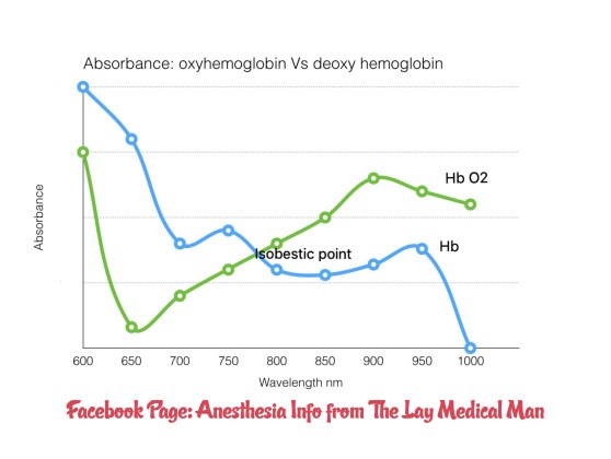
☝️️One LED emits light at 660 nm (red light) and the other at 940 nm (infrared light)

☝️️Oxyhaemoglobin and deoxyhaemoglobin absorb these wavelengths differently

☝️️Oxyhaemoglobin absorbs more infrared light (940 nm) and allows more red light (660 nm) to pass through

☝️️Deoxyhaemoglobin absorbs more red light (660 nm) and allows more infrared light (940 nm) to pass through

☝️️Isobestic point is at 806 nm

☝️️The LEDs flash in sequence: one on, then the other, then both off (to allow correction for ambient light). This triplet sequence happens 30 times per second

☝️️The amount of light transmitted through the patient at each frequency is detected by the photodiode.

☝️️The microprocessor corrects for ambient light, and also for the difference between arterial and venous saturations by deducting the minimum transmitted light, during diastole, from the maximum during systole.

☝️️After this, the ratio of oxy to deoxyhaemoglobin is determined and from this the percentage oxygen saturations is determined, using an empirical table derived from healthy volunteers who were exposed to varying degrees of hypoxia.

💅🏽Apart from the common causes like movement, nail varnish, diathermy,  others like

🔻severe anaemia

🔻cardiac arrhythmias

🔻Methaemoglobinaemia (characteristically cause saturations to be measured at around 85%)

🔻Increased venous pulsation, e.g. severe tricuspid regurgitation

🔻i.v. methylene blue dye (because it absorbs light in the 660–670 nm range also may cause erroneously low readings

💅🏻Carboxy hemoglobin (CO-Hb has similar absorption spectra as that of oxy-Hb) is detected by normal pulse oximeters as oxy hemoglobin–> erroneous high readings

💅🏻Cyanide prevents oxygen being utilised in respiration and so its extraction from the blood falls; so in cyanide poisoning, though the value is not inaccurate, it should be interpreted as inappropriately high

☝️️Fetal haemoglobin and Hb S (sickle) do not affect readings

☝️️The human volunteers used to construct empirical saturation tables did not have their oxygen saturations dropped below approximately 85%; hence readings below this number are extrapolated, not validated.

img_1994

The Life of P.I. (Perfusion Index)

🚤 Reduction of plethysmographic pulse wave amplitude (PPWA) has been proven to be a reliable method for detecting the IV injection of an exogenous vasopressor ( for e.g. The adrenaline in epidural test dose)

🚤 Currently, a numerical value has been added to new pulseoximeters indicating the PPWA, termed the perfusion index (PI), to augment its clinical applicability.

🚤i.e. PI is the numerical value of the amplitude of the plethysmographic pulse wave that is displayed on many pulse oximeters.

🚤 Using pulse oximetry, a variable amount of light is absorbed by pulsating arterial flow (AC) and a constant amount of light is absorbed by nonpulsating blood and tissue (DC). The pulsating signal indexed against nonpulsating signal and expressed as ratio is commonly referred to as the perfusion index
🚤 It depends on the distensibility of the vascular wall and the intravascular pulse pressure. Usually the effect of autonomic impulses upon distensibility is so strong that it predominates the opposite effect of pulse pressure.
🚤 Decreases in PI resulting from pain and other stressful stimuli are due to vasoconstriction of the finger arterial bed rather than changes in the pulse pressure
Reference: The Efficacy of Perfusion Index as an Indicator for Intravascular Injection of Epinephrine-Containing Epidural Test Dose in Propofol-Anesthetized Adults, Anesth Analg 2009;108:549 –53)