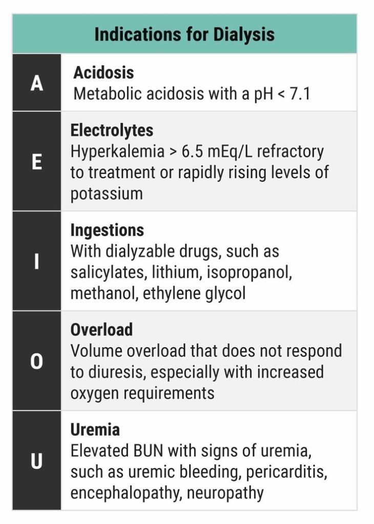
DIALYSIS INDICATIONS

ABG Exercises and Flash Cards

Arterial Blood Gas Analysis- Combined Copenhagen Physico-chemical Approach

VIVA SCENE: HYPOCALCAEMIA

Plasma calcium < 2.2 mmol/L

Normal values: Total calcium 2.25-2.60 mmol/L; ionised calcium 1.12-1.32 mmol/L

HYPOCALCAEMIA CAUSES:

  • Decreased parathyroid hormone

  • Decreased Vitamin D activity (e.g. intestinal malabsorption, liver disease, CRF)

  • Increased calcium loss (e.g. chelating agents, calcification of soft tissues)

  • Decreased ionised calcium (e.g. alkalosis)

  • Tumour Lysis Syndrome
  • Diarrhoea, vomiting, and nasogastric suction can cause hypomagnesaemia with secondary hypocalcaemia (HSH)

CLINICAL FEATURES:

  • Tetany
  • Seizures
  • Emotional instability/agitation/anxiety
  • Myopathy

ECG:

  • QTc prolongation by prolonging the ST segment

  • Torsades de pointes and atrial fibrillation in severe cases

NB: The corrected QT interval (QTc) is taken as the time between the beginning of the QRS complex and the end of the T wave, it is less than 440 ms in men and 460 ms in women. Severe hypocalcaemia (less than 1.9 mmol/L) may cause a prolongation of the QTc. A QTc greater than 500 ms is associated with an increased risk of Torsades de Pointes.

TREATMENT:

  • Ca2+ 0.5mL/kg (max 20mL) of 10% calcium gluconate OR 0.2mL/kg of 10% calcium chloride
  • Administer by slow IV (max 2 mL/min), repeat if necessary.
  • Calcium can precipitate or exacerbate digitalis toxicity therefore IV calcium must be given very slowly in patients on digoxin and the ECG must be monitored continuously

ERRORS THAT MAY HAPPEN, IF AN ARTERIAL BLOOD GAS SAMPLE REMAINS FOR QUITE SOME TIME IN ROOM AIR, BEFORE ANALYSIS

Screen Shot 2018-09-02 at 6.23.11 PM

ERRORS IN ABG SAMPLING: CAUSES & CONSEQUENCES

Screen Shot 2018-09-02 at 6.21.23 PM

pH and [H+]

Screen Shot 2018-09-02 at 5.05.35 PM