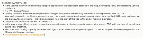
CLOSING CAPACITY
The functional residual capacity of the infant’s lungs is only one half that of an adult in relation to body weight. This difference causes excessive cyclical increases and decreases in the newborn baby’s blood gas concentrations if the respiratory rate becomes slowed because it is the residual air in the lungs that smooths out the blood gas variations.
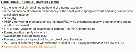
The functional residual capacity equals the expiratory reserve volume plus the residual volume. This is the amount of air that remains in the lungs at the end of normal expiration (about 2300 milliliters).


↪️Despite decreased requirements during pregnancy ,spinal anesthesia requirements return to non pregnant levels by 12-36 hours postpartum. Abouleish et al found that patients required 30% more bupivacaine, to achieve a T4 level for post partum tubal ligation , upto 24 hours after delivery. Rapid decline in plasma progesterone levels, after delivery of placenta is one factor, which contributes to this.
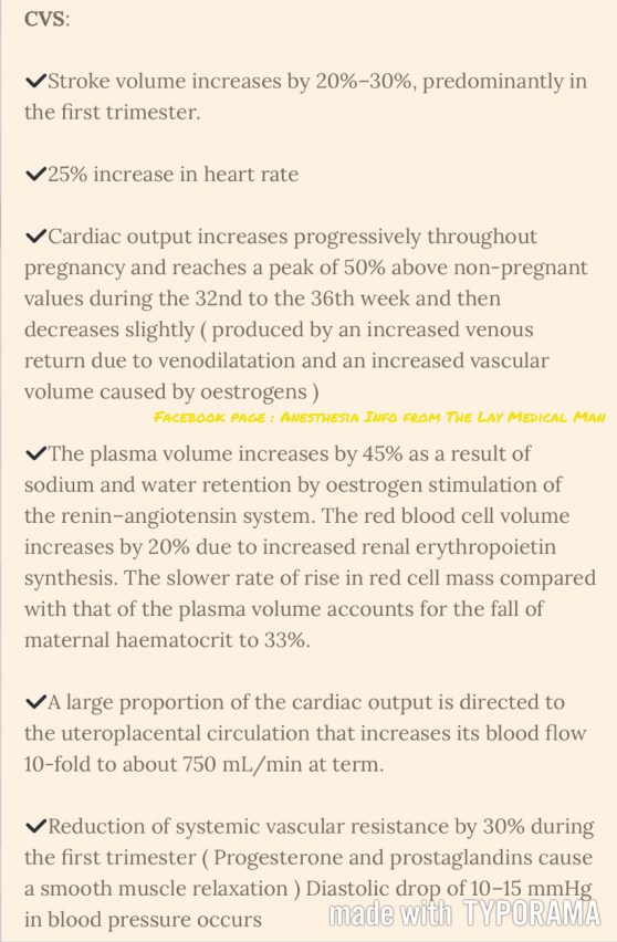
↪️Cardiac output rises immediately after delivery because of autotransfusion of 500 to 750 ml of blood from the uterus. Patients with pulmonary hypertension and stenotic valvular lesions are at a particular risk at this time.
↪️Cardiac output returns to slightly above prepregnancy values about 2 to 4 weeks after delivery.
↪️FRC and residual volume rapidly return to normal.
↪️Many of the pulmonary changes caused by mechanical compression by the gravid uterus resolve quickly. Alveolar ventilation returns to baseline by 4 weeks postpartum, and there is a rise in maternal PCO 2 as the progesterone levels decrease.
↪️The dilutional anemia of pregnancy resolves, and the hematocrit returns to normal within 4 weeks secondary to a postpartum diuresis.
↪️Serum creatinine, glomerular filtration rate, and BUN return to normal levels in less than 3 weeks.
↪️Mechanical effects of the gravid uterus on the gastrointestinal system resolve about 2 to 3 days after delivery; however, gastric emptying may be delayed for several weeks as serum progesterone levels slowly decrease.
#TubalLigation ,#pps , #PostpartumSterilization , #ObstetricAnesthesia , #ObstetricAnaesthesia, #PregnancyPhysiology ,#anesthesia , #anaesthesia ,#obstetrics , #pregnancy , #sterilization
Reference:
Shnider and Levinson's anesthesia for obstetrics, Maya Suresh; Sol M Shnider; Gershon Levinson, 2013,English : 5th
Ana M. Lobo, Andrea J. Fuller,Marina Shindell, Chapter 59, Anesthesia Secrets, 4/e


🍃Ventilatory responses to hypoxia and hypercapnia are impaired secondary to reduced central nervous system activity.
🍃The respiratory depressant effects of benzodiazepines, opioids, and volatile anesthetics are exaggerated.
🍃These changes compromise the usual protective responses against hypoxemia after anesthesia and surgery in elderly patients.
🍃The loss of elastic recoil combined with altered surfactant production leads to an increase in lung compliance.
🍃Increased compliance leads to limited maximal expiratory flow and a decreased ventilatory response to exercise.
🍃Loss of elastic elements within the lung is associated with enlargement of the respiratory bronchioles and alveolar ducts, and a tendency for early collapse of the small airways on exhalation.
🍃There also is a progressive loss of alveolar surface area secondary to increases in size of the interalveolar pores of Kohn. This results in increased anatomic dead space, decreased diffusing capacity, and increased closing capacity all leading to impaired gas exchange.
🍃Loss of height and calcification of the vertebral column and rib cage lead to a typical barrel chest appearance with diaphragmatic flattening.
🍃The flattened diaphragm is mechanically less efficient, and function is impaired further by a significant loss of muscle mass associated with aging. Functionally, the chest wall becomes less compliant, and work of breathing is increased.
🍃Total lung capacity is relatively unchanged.
🍃Residual volume increases by 5% to 10% per decade.
🍃Vital capacity decreases.
🍃Closing capacity increases with age.
🍃Functional residual capacity (FRC) is determined by the balance between the inward recoil of the lungs and the outward recoil of the chest wall. FRC increases by 1%–3% per decade because at relaxed end expiration, the rate of decrease in lung recoil with aging exceeds that of the rate of increase in chest wall stiffness.
🍃In younger individuals, closing capacity is below functional residual capacity. At 44 years of age, closing capacity equals functional residual capacity in the supine position, and at 66 years of age, closing capacity equals functional residual capacity in the upright position.
🍃When closing capacity encroaches on tidal breathing, ventilation-perfusion mismatch occurs.
🍃When functional residual capacity is below closing capacity, shunt increases, and arterial oxygenation decreases. This results in impairment of preoxygenation. Increased closing capacity in concert with depletion of muscle mass causes a progressive decrease in forced expiratory volume in 1 second by 6% to 8% per decade.
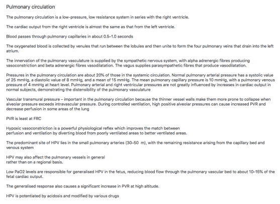
🍃Increases in pulmonary vascular resistance and pulmonary arterial pressure occur with age and may be secondary to decreases in cross-sectional area of the pulmonary capillary bed. Hypoxic pulmonary vasoconstriction is blunted in elderly individuals and may cause difficulty with one-lung ventilation.
Ref: Geriatric Anesthesia 2/e , Miller’s Anesthesia 7/e
#Physiology , #Anesthesia , #Geriatrics
PREGANGLIONIC NEURONS of the sympathetic system synapse with the POSTGANGLIONIC NEURONS in the SYMPATHETIC GANGLIA. These ganglia together will form sympathetic chain. The sympathetic chains extend down the length of the vertebral column and are divided into four parts:
CERVICAL SYMPATHETIC GANGLIA
🔸Consist of three ganglia: Superior, Middle and Inferior
🔸Superior sends postganglionic fibres to form the internal carotid plexus
🔸Inferior or Stellate ganglion is fused with the first thoracic ganglia
THORACIC SYMPATHETIC GANGLIA
🔸T1-T5 ganglia supply the aortic, cardiac and pulmonary plexus
🔸The last 7 thoracic ganglia form the greater and lesser splanchnic nerves
🔸The lowest splanchnic nerve is formed from the last thoracic ganglia and supplies the renal plexus
LUMBAR PREVERTEBRAL SYMPATHETIC GANGLIA
🔸Supplies the coeliac plexus
SACRAL SYMPATHETIC GANGLIA (PELVIC)
🔸Contribute to hypogastric and pelvic plexus
➿The autonomic nervous system is a division of the nervous system that controls the activity of internal organs.
➿The sympathetic division prepares the body for fight or flight reactions. The parasympathetic system promotes ‘rest and digest’ (restorative) functions.
➿Acetylcholine is the principal transmitter released by the preganglionic fibres of both the sympathetic and the parasympathetic nervous systems. The parasympathetic postganglionic fibres secrete acetylcholine onto their target organs, whereas norepinephrine is principally secreted by the postganglionic sympathetic fibres.
➿The central portions of the autonomic nervous system are located in the hypothalamus, brainstem and spinal cord. The limbic system and parts of the cerebral cortex send signals to the hypothalamus and lower brain centres, which can also influence the activity of the ANS
➿The posterior and lateral hypothalamic areas increase blood pressure and heart rate, whereas the preoptic area decreases blood pressure and heart rate. These effects are mediated by cardiovascular centres in the pontine and medullary reticular formation.
➿In the ANS, the connection between the CNS and its effector consists of two neurons—the preganglionic neuron and the postganglionic neuron. The synapse between these two neurons lies outside the CNS, in an autonomic ganglion [These are the cell bodies of the post ganglionic neuron, located in chains alongside the vertebral column, in plexuses in the abdomen (Sympathetic) or within the innervated target organ (Parasympathetic)]. The axon of a preganglionic neuron enters the ganglion and forms a synapse with the dendrites of the postganglionic neuron. The axon of the postganglionic neuron emerges from the ganglion and travels to the target organ #TheLayMedicalMan
➿The sympathetic system has short preganglionic fibres and long postganglionic fibres. As the parasympathetic ganglia are located near or within their effector organs, the parasympathetic postganglionic fibres are short.
➿ The pre-ganglionic fibres are slow-conducting B or C fibres. The postganglionic fibres that originate from the ganglia and innervate target organs are largely slow-conducting, unmyelinated C fibres. #TheLayMedicalMan
➿ There are more postganglionic fibres than preganglionic nerves and so the stimulation of a single preganglionic neuron can activate many postganglionic nerves, resulting in divergence. But in the superior cervical ganglion, numerous preganglionic fibres converge on a single postganglionic neuron, resulting in convergence.
Facebook page : Anesthesia Info from The Lay Medical Man
