ANTIDEPRESSANTS; AS ANALGESIC Vs AS ANTIDEPRESSANT

The tricyclic antidepressants prevent the reuptake of monoamines, including serotonin and noradrenaline, as both pathways are important in the pain propagation. So the mixed reuptake drugs work better than more selective drugs like SSRIs

But serotonin-noradrenaline reuptake inhibitor antidepressants (SNRIs), for example, venlafaxine & duloxetine and the atypical antidepressant group, such as bupropion and mirtazapine are also effective for some chronic pain conditions and are increasingly used because of their improved tolerability

The superiority of tricyclics, particularly clomipramine and amitriptyline, in the management of pain may also be explained by their additional action on sodium channels blockade ( which is an action that SNRIs do not exhibit.)

The dose of amitriptyline to treat pain is much lower when compared to that needed to treat depression

The analgesic action has a faster onset, whereas antidepressant action takes weeks to start

The sedative action of tricyclic antidepressants are helpful in treating the sleep disturbances associated with neuropathic syndromes. Nortriptyline is less sedative than amitriptyline.

#antidepressants , #analgesics , #PainManagement , #ChronicPain

Reference: Medscape, Pharmacogenetics and Analgesic Effects of Antidepressants in Chronic Pain Management, Frédérique Rodieux; Valérie Piguet; Patricia Berney; Jules Desmeules; Marie Besson, Personalized Medicine. 2015;12(2):163-175.
Ryder S A, Stannard C F. Treatment of chronic pain: antidepressant, antiepileptic and antiarrhythmic drugs. Contin Educ Anaesth Crit Care Pain 2005; 5: 18–20 .

A TRAVELOGUE: The long journey of Insulin

Insulin is produced by beta cells of islets of Langerhans.

It is produced from the pro hormone, ‘preproinsulin’ in endoplasmic reticulum. A portion of the structure is cleaved off and the remaining portion is folded with the help of C-peptide to form ‘proinsulin’

The C-peptide portion is then removed to form Insulin

This active Insulin is transported via Golgi apparatus to cytoplasmic granules for exocytosis into plasma

Insulin then binds with its receptor on Insulin sensitive cells

Insulin receptor is a tetramer consisting of 2 alpha & 2 beta units.

Insulin binds to the alpha unit on the cell membrane, while the beta unit, which spans the cell membrane activates tyrosine kinase and the second messenger system

This activates cytoplasmic vesicles containing transport molecules

The vesicles fuse with the cell membrane to incorporate the transport molecules into the cell membrane, which facilitate the transport of glucose into the cell.

MNEMO> MECHANISM OF ACTION: INSULIN Vs GLUCAGON

Insulin binding to the receptor activates an intracellular second-messenger system via tyrosine kinase.
Glucagon binding to its receptor activates a G-protein second-messenger system via adenylyl cyclase.

“Insulin is TricKy”
“Glucagon is ACcurate”

A FEW CLUES IN INTERPRETING AN ISOLATED PROLONGATION OF ACTIVATED PARTIAL THROMBOPLASTIN TIME (aPTT)

aPTT tests the intrinsic and common pathways of coagulation

Though it is included commonly as a part of coagulation profile assessment, it’s primary uses are to detect coagulation factor deficiency and titration of heparin therapy

An isolated elevation of aPTT may indicate

deficiency of Factor VIII or IX or XI or XII

acquired clotting factor inhibitors

presence of Lupus anticoagulant

N.B.:- Factor VIII deficiency is Haemophilia A, Factor IX deficiency is Haemophilia B and Factor XI deficiency is Haemophilia C

If factor levels are >30% of normal, aPTT may remain normal, for e.g. in mild von Willebrand disease [raised aPTT + prolonged Bleeding Time (BT)], in mild hemophilia etc

Reference: Martlew V. Peri-operative management of patients with coagulation disorders. Br J Anaesth. 2000; 85(3): 446–455.

HOMOCYSTINURIA : Anesthesia IMPLICATIONS

There is increased levels of homocystine and methionine in blood and urine due to the deficiency of Cystathionine B synthetase which catalyses the conversion of homocystine and serine into cystathionine

Raised cystine levels reduce the resistance of endothelium against thrombosis, reduces the activity of the vasodilator nitric oxide (NO) and increase platelet aggregation. So there is high incidence of thromboembolism. We have to ensure good hydration, good cardiac output,early mobilisation and should provide mechanical +/- pharmacological thromboprophylaxis. Many patients will be on anticoagulation. If untreated 50% of patients will have thromboembolic complications and the mortality is about 20% before the age of 30 years. So both modification of the dosing of anticoagulants ( especially if regional anesthesia is planned) if patient is receiving them and providing prophylaxis against DVT are important elements of perioperative care. The incidence of thrombotic complications are more in pregnant patients.

Blood viscosity and platelet adhesiveness can be reduced by dextran, and the prior administration of pyridoxine

Reduced cystine results in weak collagen and fragmentation of elastic tissue of large arteries. There is high incidence of vascular diseases like Cerebrovascular diseases, Coronary Artery Disease, Peripheral Vascular Diseases

Patients may have increased insulin levels resulting in hypoglycemia. Dextrose infusion will prevent hypoglycaemia.

Acute psychiatritc symptoms, delirium etc have been reported and the altered availability of homocysteine, methionine and cystiene which are having glutamate agonist properties, has been postulated as a factor which promotes this.

Regional anaesthesia has certain theoretical disadvantages. Penetration of a large epidural blood vessel might initiate thrombosis, as may the accompanying venous stasis of the lower limbs.

Reference: ANAESTHESIA DATABOOK, A Perioperative and Peripartum Manual, 3RD EDITION
Rosemary Mason

A FEW FACTS ABOUT COAGULATION FUNCTION, IT’S MONITORING & Regional Anesthesia IN OBSTETRIC PATIENTS

During routine epidural or spinal anaesthesia, accidental puncture of epidural veins occurs in 1–18% of patients

The incidence of hematoma after epidural techniques is estimated to be in the order of 1:150,000 after epidural placement and 1:220,000 after spinal injection in the general population

removal of epidural catheters posed an equal risk to insertion ( Van- dermeulen et al)

Surgery on spinal haematoma should ideally be performed within 8–12 h of the identification of symptoms in order to improve the chances of recovery.

The overall risk of death in those having general anaesthesia for caesarean section was quoted in 2007 as being just over 1:25,000.

The levels of factors VII, VIII and fibrinogen increase and those of anticoagulation factors decrease, causing augmented coagulation and decreased fibrinolysis in pregnancy.

There is no evidence to support routine full blood count (FBC) or coagulation tests in women before the performance of a regional block in those who have had

normal FBC results

no bleeding history

no signs or symptoms of liver disease

no signs or symptoms of pre-eclampsia, abruption or clinical signs of disseminated intravascular coagulation

no recent anticoagulant treatment.

In women with known thrombocytopaenia, a Full Blood Count (FBC) should be checked within 24 h of a regional procedure.

In women with mild to moderate pre-eclampsia, the course of the disease can be unpredictable and so FBC be checked within 6 h. In addition, coagulation tests should be performed if platelets are <100000/mcL or if there is abnormal liver function.

In severe disease, FBC and clotting should be checked immediately before a procedure, as platelet levels in particular can decline rapidly.

Women with pregnancy-induced hypertension alone do not require an FBC before a regional procedure

Activated partial thromboplastin time ratio (APTTR) and international normalised ratio (INR) are slightly decreased in late pregnancy.

In a patient who receives LMWH, if he/she is simultaneously taking NSAID+Aspirin, there is an increased risk if last dose of LMWH is between 12-24 hours; it further increases if last dose is <12 hours

In patients with pre-eclampsia and platelet count between 75000-100000/mcL, there is an increased risk even if coagulation tests are normal; but it increases further if the counts has not been stable (=decreasing platelet count)

#obstetrics , #anesthesia , #coagulation , #anaesthesia

Reference: Abnormalities of Coagulation and Obstetric Anaesthesia, Hilary Swales, AAGBI Core Topics in Anaesthesia 2015

VIVA SCENE: COMPATIBILITY IN BLOOD TRANSFUSIONS: RBC Vs FFP Vs PLATELETS AND OTHER QUESTIONS

COMPATIBILITY: RBC TRANSFUSION

In red cell transfusion, there must be ABO and RhD compatibility between the donor’s red cells and the recipient’s plasma.

All healthy normal adults of group A, group B and group O have ANTIBODIES IN THEIR PLASMA against the red cell types (antigens) that they have not inherited

Among the ABO blood groups:

Group A individuals have antibody to group B

Group B individuals have antibody to group A

Group O individuals have antibody to group A and group B

Group AB individuals do not have antibody to group A or B. So,

1 Group O individuals can receive blood from group O donors only ( as the antibodies against A or B in their plasma will react with any A or B antigens which enter the circulation)

2 Group A individuals can receive blood from group A and O donors

3 Group B individuals can receive blood from group B and O donors

4 Group AB individuals can receive blood from AB donors, and also from group A, B and O donors ( as their plasma don’t have any antibodies against any antigens)

RhD RED CELL ANTIGENS AND ANTIBODIES

Is the second most important group system. Out of the existing C,D and E antigens, D is the most antigenic one. Anti D antibodies are not normally found in the blood of Rh negative individuals; instead they develop it only when itcomes into contact with Rh positive blood during child birth or inappropriate transfusion. In case of subsequent transfusins or pregnancies with Rh positive blood- this can cause rapid destruction of RhD positive red cells (Hemolytic disease of the newborn[HDN] in subsequent pregnancies; to prevent this sensitization we should give Rhesus imunoglobulin= Anti-D prophylaxis- to the Rh negative mother who gave birth to an Rh positive baby). The fetal red cells are haemolysed, causing severe anaemia. HDN due to ABO incompatibility is usually less severe than Rh incompatibility.). FFP does not need to be Rh-compatible. Anti-D prophylaxis is not necessary in Rh D-negative recipients of Rh D-positive FFP. 

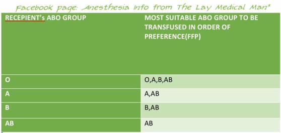
PLASMA TRANSFUSION: COMPATIBILITY

In plasma transfusion, group AB plasma can be given to a patient of any ABO group because it contains neither anti-A nor anti-B antibody.

1 Group AB plasma (no antibodies) can be given to any ABO group patients

2 Group A plasma (anti-B) can be given to group O and A patients

3 Group B plasma (anti-A) can be given to group O and B patients

4 Group O plasma (anti-A + anti-B) can be given to group O patients only

FFP does not need to be Rh-compatible (However, the unit will still be labelled as Rh +ve or Rh −ve) ; anti-D prophylaxis is not necessary in Rh D-negative recipients of Rh D-positive FFP

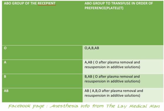
PLATELET TRANSFUSION: COMPATIBILITY

The Platelet Concentrates( PCs ) transfused must be ABO-identical, or at least ABO-compatible, in order to give a good yield ( Ideally, ABO identical units should be used but, in an emergency, ABO non-identical units can be used, although the improvement seen in platelet count post-transfusion may be less.)

Group O PC can be used for patients with blood groups A, B, and AB ONLY IF, they are resuspended in additive/preservative solutions, or if negative for high titre anti-A/A,B

ABO-incompatible PCs have reduced efficacy and, preferably, should not be used

Rh-negative patients, in particular women of childbearing age, should receive, if possible, RhD-negative PC

In the case of a transfusion of a RhD-positive PC to a RhD-negative women of childbearing age, 250 IU (50 μg) of anti-D immunoglobulin should be administered, a dose able to cover the transfusion of five therapeutic doses of PC in 6 weeks

ACUTE EMERGENCY : COMPATIBILITY

During an acute emergency, the blood bank may send group O (and possibly RhD negative) blood, especially if there is any risk of errors in patient identification. This may be the safest way to avoid a serious mismatched transfusion, in such situations.

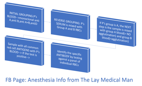
HOW A GROUP AND SAVE IS PERFORMED? (P’s = Patient’s)

Screen Shot 2019-08-11 at 11.48.54 am

#BloodTransfusion , #ABO , #BloodGroup , #TransfusionMedicine , #Anaesthesia , #Anesthesia , #Bloodbank

Reference: The Clinical Use of Blood, Handbook, WHO,
Recommendations for the transfusion of plasma and platelets Giancarlo Liumbruno, Francesco Bennardello, […], and as Italian Society of Transfusion Medicine and Immunohaematology (SIMTI) Working Party

15000768_1139329719436181_1357356490299776143_o14915305_1139329722769514_5358933164978110125_n


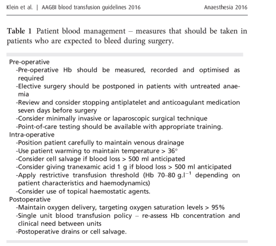
AAGBI GUIDELINES 2016

Screen Shot 2019-08-11 at 12.01.43 pm

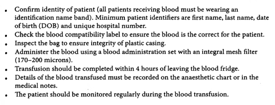
HOW WILL YOU TRANSFUSE THE BLOOD?

Screen Shot 2019-08-11 at 1.16.04 pm

Also preoperatively the need for transfusion must have been explained and written informed consent should have been taken

A general Hb threshold of 7.0 g/dl should apply as a guide for red cell transfusion. 8.0 g/dl for patients with IHD

ALSO NOTE:

A transfusion of 10 ml/kg of RBC should increase Hb by approximately 2.0 g/dl-

Cryoprecipitate should be given in a dose of 510 ml.kg-1

Platelets should be given in a dose of 1020 ml.kg-1.

Fresh frozen plasma may be given in doses of 1015 ml.kg-1.

Tranexamic acid can be used in children: a loading dose of 15 mg.kg-followed by infusion 2 mg.kg-1.h-1 should be used in trauma

Succinylcholine aka Suxamethonium

Two molecules of acetyl choline joined together by the acetyl group forms Succinylcholine

It can be presented as chloride, bromide or iodide salt

When presented as the chloride salt, it’s a solution with concentration 50 mg/ mL

When presented as bromide or iodide salts, they are powders, with more stability, shell life and suited for warm climates; but has to be reconstituted before use

pH of the solution is around 4

So they are destroyed by mixing it with alkaline solutions (e.g. Thiopentone )

#anesthesiology , #anaesthesia , #pharmacology

Reference: Kestin I. Suxamethonium. Update in Anaesthesia 1992; 1: article 7. Peck T, Hill S, Williams M. Pharmacology for Anaesthesia and Intensive Care, 3rd edn. Cambridge: Cambridge University Press, 2008; pp. 179–84 .

Pharmacological factors determining the quality of epidural blockade when using a continuous local anesthetic infusion

14522753_1129522723750214_2057384185164985570_n

FIBROMYALGIA- AN OVERVIEW

Is a common chronic pain condition, characterised by

Pain ( Spontaneous, widespread , diffuse, worse in the morning, hypersensitivity to all painful stimuli, >3 months duration, 11 out of 18 defined tender points produce tenderness on digital palpation)
Sleep disturbances
Fatigue

Pathophysiology may include

dysfunction of descending inhibitory pathways
abnormal neurotransmitter release
central sensitisation etc

Tricyclic antidepressants ( like Amitriptyline 5-10 mg ) may be effective in fibromyalgia as they reduce pain & fatigue and improve sleep

Other therapies used:

Pregabalin
Gabapentin
Newer MAO inhibitors like pirlindole
TENS
Acupuncture
Intravenous lignocaine
Injection of trigger points
Cognitive Behavioural Therapy
Warm bath
Complimentary therapies

#pain , #fibromyalgia , #PainManagement

Reference: Dedhia JT, Bone ME. Pain and fibromyalgia. Contin Educ Anaesth Crit Care Pain. 2009; 9(5): 162–166.

PIN INDEX

14721733_1121864291182724_3787877376021354340_n