LUMBAR PLEXUS BLOCK

Anatomy of lumbar plexus?

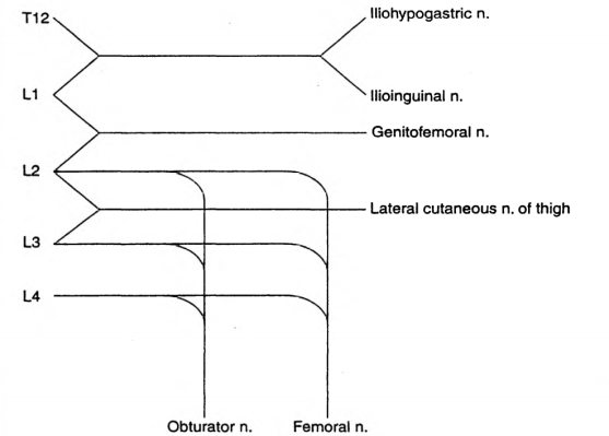
The lumbar plexus is formed by the anterior primary rami of L1 to L4 (Sometimes T12 also may contribute)
L1 forms the iliohypogastric and ilioinguinal nerves
It also gives a contribution to the formation of the genitofemoral nerve
L2 forms the lateral cutaneous nerve of thigh, along with L3
The obturator and femoral nerves are formed by contributions from L2,L3,L4

What are the indications?

Surgeries involving hip/thigh/upper leg/trauma
In conjunction with sciatic nerve or sacral plexus block
Cancer pain arising from hip or upper femur
Sympathetic block also helps in ischemic pain and CRPS

Which approach will you use?

I will use the 3 in 1 approach that aims to block the femoral, obturator and lateral cutaneous nerves with a low anterior approach. The patient is in the supine position.I will use a nerve stimulator. Using a 50 mm insulated needle, I will puncture the skin at a point 1 cm lateral to the femoral pulse and 2 cm below the inguinal ligament, at 45 degrees to skin and directed proximally and parallel to the femoral artery. The endpoint is a quadriceps twitch, which occurs at a depth of 30-50 mm. I will press distal to the injection to enhance the spread of the drug proximally. I will block the lateral cutaneous nerve separately by injecting 10 ml of local anesthetic at a point 2 cm inferior and medial to the ASIS. Other approaches are psoas compartment block and fascia iliacus block

How does the lumbar sympathetic block differ from the lumbar plexus block?

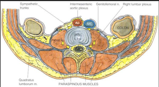
The lumbar sympathetic nerves lie anterolateral to the vertebral body, whereas the somatic nerves lie posterior to the psoas muscle and fascia. The sympathetic chains have anteriorly, aorta on the left and IVC on the right. The aim is to deposit local anesthetic around the nerves from L2 to L4 either with a single injection at L3 or 3 separate injections at L2, L3 and L4. It is used in lower limb ischemia, CRPS, phantom limb, urogenital pain etc

How to do the lumbar sympathetic block?

Patient should be positioned lateral with the side to be blocked up. A point is marked 8 cm from the midpoint of the spinous process of the desired vertebra. Its done under image guidance. A 12 cm 22 G needle is inserted at 45 degree angle directed medially towards the vertebral body which lies at a depth of around 8 cm; if the needle hits the bone at 4-5 cm depth it is likely to be the transverse process and should be redirected cranially or caudally to pass over it. Once the needle hits the vertebral body, it should be redirected slightly antero-laterally till we feels the pop-off of passing the psoas fascia. Then the local anesthetic mixed with radiographic contrast is injected which should form a band around the desired vertebral bodies. If the contrast disappears very quickly, it may be in a vessel. Otherwise, it can go into the psoas muscle or retroperitoneal tissue. Also we should take lateral and anteroposterior images to confirm the correct position.

Complications of lumbar sympathetic block?

Local anesthetic toxicity due to injection into aorta or IVC
Profound motor block or permanent paralysis due to intrathecal injection
Profound hypotension: good iv access and access for resuscitation equipments are a must
Post sympathectomy pain
Ureteric injury, ejaculatory failure

Screen Shot 2019-03-18 at 3.15.22 PMScreen Shot 2019-03-18 at 11.01.44 AM

MONITORING OF NEUROMUSCULAR BLOCKADE

  • ASSESSMENT OF NEUROMUSCULAR FUNCTION
  • CLINICAL:
  • Grip strength
  • Ability to sustain head lift for at least 5 seconds
  • Ability to produce vital capacity of at least 10 mL/kg
  • NEUROMUSCULAR STIMULATION EQUIPMENTS:
  • Peripheral nerve stimulator
  • Mechanomyography: uses force transducer to quantitatively measure contractile response
  • Acceleromyography: measures movement of joints caused by muscle movement
  • Electromyography: measures electrical activity associated with action potential propagation in a muscle cell (research use)
  • BENEFITS
  • Monitoring helps to assess the degree of relaxation, help adjust dosage, assess development of phase II block, provide early recognition of patients with abnormal cholinesterases, and to assess cause of apnoea.
  • All twitches that we deliver in theatre with the nerve stimulators are uniform
    and so we only need to learn a couple of facts.
    • All twitches are delivered at 50 mA (i.e. supramaximal stimulus)
    • All twitches last 0.2 ms
    Once we have these facts, the rest becomes easier to remember
  • TYPES
  • Single twitch is insensitive since >75% of postsynaptic receptors must be blocked before there is any diminution in twitch height. We will not see ‘fade’ during single-twitch stimulation. >> Twitch current 50 mA. Duration of twitch 0.2 ms. Frequency of twitches 1 Hz (i.e. 1 every second). Number of twitches: as many as operator chooses to give. Single twitch can be used to assess block in depolarising nmbd, i.e.
    suxamethonium, where fade and post-tetanic facilitation do not occur
  • Train of four: >>Twitch current 50 mA. Duration of twitch 0.2 ms. Frequency of twitches 2 Hz (i.e. 2 every second). Number of twitches: 4. Compare last (T4) and first twitch (T1). TO4 ratio (T4 :T1) indicates degree of neuromuscular blockade:
  • – T 4 disappears at 75% depression of T 1 (1st, 2nd and 3rd twitches present)
  • – T 3 disappears at 80% depression of T 1 (1st and 2nd twitches present)
  • – T 2 disappears at 90% depression of T 1 (1st twitch only)
  • – T 1 disappears at 100% depression of T 1 (no twitches).
  • A TO4 count of 0–1 is needed for adequate intubating conditions, but a count of three twitches provides adequate relaxation for most surgery (To4 ratio of 0.15 – 0.25) To4 ratio of > 0.9: essential for safe extubation and recovery post surgery
  • A TO4 ratio >0.70, corresponds to adequate clinical recovery, but normal pharyngeal function requires a ratio >0.90.
  • Neuromuscular reversal can be given when T2 has reappeared, i.e. when T1 is about 20% of its control height.
  • The diaphragm is the most resistant (but with shorter onset times) of all muscles to both depolarising and non-depolarising relaxants requiring 1.5 to 2 times as much drug as the adductor pollicis muscle for an identical degree of blockade.
  • A supramaximal stimulus should be truly maximal throughout the test period to maintain accuracy; hence the electrical current applied is at least 20% to 25% above that necessary for a maximal response.
  • Tetanic stimulation: >> Twitch current 50 mA. Duration of twitch 0.2 ms. Frequency of twitches 50 Hz (i.e. 50 every second). Number of twitches: stimulation lasts 5 seconds = 5 × 50 = 250 twitches. With non-depolarizing block, peak height is reduced and fades. Release of acetylcholine is reduced (possibly presynaptic effect) and postsynaptic receptors are blocked, limiting sustained contraction. Tetanic stimulation is extremely painful in an awake patient and may leave an unpleasant sensation in those who were anaesthetised.
  • Post-tetanic count: >> Twitch current 50 mA. Duration of twitch 0.2 ms. Frequency of twitches 1 Hz (i.e. 1 every second). Number of twitches: as many as operator chooses to give. May result in response (post-tetanic potentiation), even if none is seen with original TO4. Due to increased synthesis and mobilization of acetylcholine following tetanus. Appearance of post-tetanic count precedes return of TO4 by 30–40 min.PTC < 5 = profound block. PTC >15 = equivalent to two twitches on To4
  • Double-burst stimulation: >> Twitch current 50 mA. Duration of twitch 0.2 ms. Frequency of twitches 50 Hz (i.e. 50 every second). Number of twitches: 3 twitches – break of 750 ms – 3 more twitches. Similar to TO4, but tactile evaluation is more sensitive because fade of the two resultant contractions is more marked.
  • Nondepolarising neuromuscular blocking agents (NDMB): repetitive stimulation (ToF or tetanus) is associated with fade (reduction in amplitude of evoked responses with T4 affected first, then T3, followed by T2, then finally T1) and post-tetanic facilitation.
  • Depolarising neuromuscular blocking agents (DMB): no fade or posttetanic facilitation observed. Repeated dose of suxamethonium can give characteristics of NDMB—phase II block).
  • A PNS is Portable, battery-powered, and easy to use Able to deliver different impulses. Supramaximal current output of 50–60 mA at all frequencies can be given to ensure all nerve fibres are depolarised. Has a Monophasic square waveform. Can give Single twitch at 0.1 Hz, Train of four (TOF) at 2 Hz and Tetanic stimulation at 50 Hz
  • Fade: A gradual diminution of evoked response during prolonged or repeated nerve stimulation, is indicative of a nondepolarizing block. Adequate clinical recovery correlates well with the absence of fade.
  • PHASE I and PHASE II  Blocks: These terms refer to the blocks seen following the administration of suxamethonium. A phase I block describes the block seen following the administration of a single dose of suxamethonium. Suxamethonium binds to the ACh receptor, which causes opening of the sodium channel and membrane depolarisation. This results in disorganised muscle contraction, seen as fasciculation followed by flaccid paralysis because suxamethonium causes prolonged depolarisation of the motor end plate. The characteristics of a phase I block:

• Reduced twitch height, but sustained response to tetanic stimulation
• No post-tetanic facilitation; does not exhibit fade during tetanus or train-of-four

• TOF ratio >70% (height of fourth twitch to that of first). This is a measure of the pre-synaptic effect of suxamethonium.
The block is potentiated by the effect of anticholinesterases because these
will further decrease the rate of suxamethonium breakdown.
A phase II block describes the block seen following the repeated administration/infusion of suxamethonium and can develop with doses
in excess of 2.5 mg/kg. It occurs because in the continued presence
of suxamethonium, the receptors eventually close and the membrane
repolarises, at least partially. However, it is now desensitised to ACh and so
cannot open again to propagate an action potentials. In this way, a phase II
block is similar to a non-depolarising block. Phase ii blocks are also called
‘desensitisation blocks’.
characteristics of a phase ii block:
• Exhibits fade on tetanic stimulation
• Exhibits post-tetanic facilitation
• TOF ratio < 0.3 (fourth to first twitch height)
• Antagonised by anticholinesterases
• Tachyphylaxis is seen with the need to increase suxamethonium infusion
rate or bolus dose.

  • Post-tetanic potentiation: Tetanic stimulation is a supra-maximal stimulation, applied to the NMJ for a prolonged period of time. It is sufficient to produce a substantial increase in ACh release, enough to overcome competition from NMBA in all but the most profound of blocks. The positive feedback mechanism via prejunctional receptors by stimulating an increase in ACh production by second messenger systems gets activated and this increases the amount of ACh available for release. This is called post-tetanic potentiation.
  • Commonly monitored nerves in theatre are:
    • Facial nerve – twitch of the eyebrow with orbicularis oculi contraction
    • Ulnar nerve – twitch of the thumb with adductor pollicis contraction
    • Posterior tibial nerve – twitch of the big toe with flexor hallucis brevis contraction
  • Which muscles are affected first by NMBAs?
    NMBAs cause paralysis of all voluntary muscles in the body, but some are
    more sensitive than others. in order of decreasing sensitivity:
    • Eyes (affected first)
    • Facial muscles
    • Neck
    • Extremities
    • Limbs
    • Abdominal muscles
    • Glottis
    • Intercostal muscles (affected last).
    Muscle function returns in the reverse order. This is why it is traditional to
    wait until a patient can lift their head of the pillow before extubation

ANTICOAGULANTS AND BRIDGING

N.B. Corrections:

LMWH inhibits factor Xa (Not XIa)

REMIMAZOLAM: IMPORTANT POINTS

  • Its an ultra-short acting benzodiazepine that is currently under investigation in phase II and III clinical trials.
  • It is metabolized by tissue esterases in the blood to its inactive metabolite, carboxylic acid, which allows for rapid removal of the drug even with its use in prolonged infusions.
  • Remimazolam is eliminated by first-order pharmacokinetics, therefore with prolonged infusions or high doses, there is no problem with drug or metabolite accumulation .
  • It has been evaluated as a premedication drug prior to anesthesia; however due to its very short duration of action and the fact that it is not available as an oral formulation limits its use in this clinical situation.
  • It has been most studied as a drug for use in procedural sedation. It has been studied widely in sedation for endoscopic procedures like colonoscopies. Studies have shown doses of 0.1-0.2 mg/kg were effective sedative doses for patients undergoing endoscopic procedures. It allows for a faster onset and recover time when compared with midazolam. There is limited incidence of respiratory depression and it canability to be reversed with flumazenil without resedation
  • It is also currently being studied as a general anesthetic, using induction doses of 6 and 12 mg/kg/h and maintenance rates of 1 mg/kg/h.
  • Because its metabolism is organ-independent, it also has been evaluated for use as a sedative agent in the ICU setting. Its ultra-short acting nature would also make it an ideal agent to allow for neurological evaluation very soon after an infusion has been stopped .
  • The safety profile of remimazolam has been shown to be favorable overall, demonstrating less vasopressor use compared with patients sedated with propofol

ETOMIDATE- BAD ‘WOW’ FACTORS

  • While it continues to be used infrequently in the UK it has been withdrawn in North America and Australia
  • The most notable and potentially serious side effect of etomidate administration is the suppression of adrenocortical steroid synthesis.
  • It suppresses adrenocortical function by inhibition of the enzymes 11-hydroxylase and 17-hydroxylase, resulting in inhibition of cortisol and aldosterone synthesis.

  • After a single bolus dose of etomidate, this adrenocortical suppression lasts approximately 6 h in healthy individuals. However in the critically ill, such suppression can last for days. In other words the situation in which it has the best cardiovascular proile is the unwell patient in whom the consequences of steroid inhibition are likely to be the most detrimental.
  • Etomidate is approximately 100-fold more potent a suppressor of adrenocortical function than it is a sedative-hypnotic. Consequently, an anesthetic
  • induction dose of etomidate represents a massive overdose with respect to its ability to suppress adrenocortical function. And etomidate s terminal elimination half-life is rather long. Thus, after just a single anesthetic induction dose of etomidate, many hours must pass before etomidate s concentration in the blood falls below that which suppresses adrenocortical steroid synthesis.

  • It is within this mechanistic context that the strategy emerged to design analogues of etomidate

  • ANALOGUES:
  • MOC-etomidate [ relatively low potency and very rapid metabolism (1.) required the administration of extremely large doses]
  • CPMM etomidate [ it has an onset and offset of hypnotic action that are fast (1.) ]
  • Carbo etomidate [ less adrenocortical inhibition (2.)]
  • MOC-carboetomidate [ combines properties (1) and (2); but it’s potency
  • is very low which means that extremely large doses would need to be administered to maintain anesthesia ]

  • Involuntary movements (myoclonus) are commonly observed after etomidate administration, with some studies reporting an incidence as high as 80 % in unpremedicated patients

  • It has been suggested that it occurs because etomidate depresses inhibitory neural circuits in the central nervous system sooner and at lower concentrations than excitatory circuits.
  • Regardless of the mechanism, myoclonus can be significantly reduced or completely prevented by administering a variety of drugs with central nervous system depressant effects including opiates, benzodiazepines, dexmedetomidine, thiopental, lidocaine, and magnesium.

  • Pain at the injection site is another common side effect and its incidence is highly dependent upon the size of the vein into which it is injected and the formulation that is used.

  • Lipid emulsion and cyclodextrin formulations may reduce TRP channel activation, leading to less pain on injection

  • Postoperative nausea and vomiting is common with reported incidences as high as 40 %. It has been suggested that the emetogenic trigger in etomidate is the propylene glycol solvent and not the anesthetic itself.

  • Reference: Ref: Pharmacology for Anaesthesia and Intensive Care, Peck and Hill, 4/e, p:105, Total Intravenous Anesthesia and Target Controlled Infusions, A comprehensive global anthology, Anthony R Absalom

  • Screen Shot 2018-10-02 at 9.11.39 PM

CONTEXT SENSITIVE HALF TIME [CSHT]

Screen Shot 2018-09-21 at 11.24.29 AM

  • Context sensitive half-time is deined as the time for the plasma concentration to fall to half of the value at the time of stopping an infusion

  • The half time will usually alter in the setting of varying durations  of drug infusion

  • The higher the ratio of distribution clearance to clearance due to elimination, the greater the range for context-sensitive half-time
  • The longest possible context-sensitive half-time is seen when the infusion has reached steady state, when there is no transfer between compartments and input rate is the same as elimination rate
  •  Draw and label the axes; draw the curve for the drug with the shortest CSHT first before plotting the others
  • REMIFENTANIL: Here the elimination always dominates distribution and so there is very little variation in CSHT with time and so it is context insensitive. Draw a straight line starting from the origin and becoming near horizontal after the CSHT reaches 5 min. This demonstrates that the half time is not dependent on the length of infusion as clearance by plasma esterases is so rapid. For remifentanil the

    longest possible CSHT is only 8 minutes

  • PROPOFOL: For propofol the clearance due to elimination is similar to that for distribution into the second compartment, so plasma concentration falls rapidly after a propofol infusion mainly due to rapid elimination with a smaller contribution from distribution. Propofol is not context insensitive as its CSHT continues to rise; however it remains short even after prolonged infusions. Starting at the origin, draw a smooth curve rising steadily towards a CSHT of around 40 min after 8 h of infusion.
  • ALFENTANIL: The curve rises from the origin until reaching a CSHT of 50 min

    at around 2 h of infusion. Thereafter the curve becomes horizontal. This shows that alfentanil is also context insensitive for infusion durations of 2 h or longer

  • THIOPENTONE SODIUM: The curve begins at the origin but rises more steeply than the others so that the CSHT is 50 min after only 30 min infusion duration. The

    curve should be drawn like a slightly slurred build-up exponential reaching a CSHT of 150 min after 8 h of infusion. As the CSHT continues to rise, thiopental does not become context insensitive

  • FENTANYLThe most complex curve begins at the origin and is sigmoid in shape. It should cross the alfentanil line at 2 h duration and rise to a CSHT of 250 min after 6 h of infusion. Again, as the CSHT continues to rise, fentanyl does not become context insensitive.

  • The maximum possible CSHT for propofol is about 20 minutes, compared with 300 minutes for fentanyl

  • It is important to realize that the CSHT does not predict the time to patient awakening but simply the time until the plasma concentration of a drug has fallen by half. The patient may need the plasma concentration to fall by 75% in order to awaken, and the time taken for this or any other percentage fall to occur is known as a decrement time.

  • Decrement time: The time taken for the plasma concentration of a drug to fall to the specified percentage of its former value after the cessation of an infusion designed to maintain a steady plasma concentration (time). The CSHT is, therefore, a form of decrement time when the specified percentage’ is 50%.

  • Although the CSHT for propofol has a maximum value of about 20 minutes, during long, stimulating surgery infusion rates will have been high and the plasma concentration when wake-up is required may be very much less than half the plasma concentration at the end of the infusion. Thus time to awakening using propofol alone may be much longer than the CSHT. This is why the TCI pumps display a decrement time rather than a CSHT.

  • When using propofol infusions, the decrement time is commonly quoted as the time taken to reach a plasma level of 1.2 μ g.ml1 , as this is the level at which wake up is thought likely to occur in the absence of any other sedative agents.

  • It must be remembered that after one CSHT, the next period of time required for plasma concentration to halve again is likely to be much longer. This relects the increasing importance of the slower redistribution and metabolism phases that predominate after re-distribution has taken place. This explains the emphasis on half-time rather than halflife: half-lives are constant whereas half-times are not!

MEDIASTENAL TUMOURS & THE ANESTHESIOLOGIST: SPECIFIC POINTS

  • A preoperative CT scan will show the site, severity, and extent of the airway compromise to assess the level and degree of obstruction.
  • Assess the vocal cord function preoperatively
  • Lung function tests to look for the extent of intrathoracic or extrathoracic obstruction.
  • ECHO to rule out pericardial effusion and cardiac compression.
  • Premedication with benzodiazepine is generally avoided if there is risk of airway compromise.
  • Airway equipment—rigid bronchoscopy and difficult airway trolley, jet ventilation, cardiopulmonary bypass (CPB) should be there as standby. Femoro femoral bypass is the most common setup.
  • COMPLICATIONS DUE TO MASS EFFECT OF THE TUMOUR:
  • Vascular compromise—SVC Obstruction ( SVCO ) and pulmonary vessel obstruction
  • Laryngeal nerve palsy
  • Dysphagia
  • STRIDOR and airway compromise may be an important symptom
  • Inspiratory stridor (laryngeal)—obstruction above the level of glottis
  • Expiratory stridor (tracheobronchial)—obstruction in the intrathoracic airways
  • Biphasic stridor—obstruction between glottis and subglottis or a critical obstruction at any level
  • Sometimes you may have to go for a microlaryngoscopy tube (MLT)
  • TAKE CARE:
  • Aim to avoid worsening of cardiac compression, airway occlusion, and SVC obstruction.
  • IV cannula in the lower extremity
  • Induction in sitting position (semi Fowler’s position)
  • Inhalational (preferred choice) or IV induction agent titrated to effect
  • Choose spontaneous ventilation with LMA
  • Awake fibreoptic technique if intubation is necessary with a reinforced smaller calibre and longer endotracheal tube
  • Postoperative airway obstruction due to airway oedema, tracheomalacia, and bleeding warrant the need for awake extubation in ITU. The following steps would aid in an uneventful extubation:
  • Test for leak around the endotracheal tube cuff.
  • Administer dexamethasone or chemo radiotherapy in sensitive tumours to shrink size of tumour.
  • Use adrenaline nebulisers.
  • Extubate over airway exchange catheters.
  • SVCO: challenges during anaesthesia
  • Need for supplemental oxygen
  • Orthopnoea—induction in the sitting-up position
  • IV cannula in the lower extremity
  • Airway oedema
  • Mucosal bleeding
  • Laryngeal nerve palsy
  • Haemodynamic instability due to decreased venous return
  • OTHER CONCERNS
  • General anaesthesia, causes loss of intrinsic muscle tone, decreased lung volumes, and decreased transpleural pressure gradient
  • Positive pressure ventilation, can precipitate severe hypotension and also increases intrathoracic tracheal compression
  • Coughing, as it can cause complete airway obstruction by positive pleural pressure, increasing intrathoracic tracheal compression
  • Following gas induction, the patient stops breathing and if you are unable to ventilate her: Follow difficult or failed intubation guidelines. But cricoid puncture and emergency tracheostomy are futile if the level of airway obstruction is at the intrathoracic tracheobronchial tree: Try a change in position—lateral, sitting up, or prone—to decrease the mechanical effect of the tumour. Avoid positive pressure ventilation for fear of luminal closure. Low-frequency jet ventilation with Sander’s injector or high-frequency translaryngeal jet ventilation with Hunsaker’s catheter is one option. CPB bypass and ECMO to restore oxygenation when other measures fail.
  • Following chemotherapy in ICU, if patient develops hyperkalemia, Tumour Lysis Syndrome should be there in the differential diagnosis
  • ALSO NOTE
  • During inspiration, the intrathoracic airways expand along with the expanding lungs. In contrast, the extrathoracic airways diminish in caliber during inspiration due to their intraluminal pressure being lower than the atmospheric pressure. The reverse happens during expiration.
  • Flow volume loop in upper-airway obstruction:
  • Fixed lesions [extrathoracic or intrathoracic] are characterized by lack of changes in caliber during inhalation or exhalation and produce a constant degree of airflow limitation during the entire respiratory cycle. Its presence results in similar flattening of both the inspiratory and expiratory portions of the flow-volume loop
  • Variable lesions are characterized by changes in airway lesion caliber during breathing. Depending on their location (intrathoracic or extrathoracic), they tend to behave differently during inhalation and exhalation.
  • In the case of an extrathoracic obstructing lesion, during inspiration, there is acceleration of airflow from the atmosphere toward the lungs, and the intraluminal pressure decreases with respect to the atmospheric pressure due to a Bernoulli effect, resulting in the limitation of inspiratory flow seen as a flattening in the inspiratory limb of the flow-volume loop. During expiration, the air is forced out of the lungs through a narrowed (but potentially expandable) extrathoracic airway. Therefore, the maximal expiratory flow-volume curve is usually normal.
  • Variable intrathoracic constrictions expand during inspiration, causing an increase in airway lumen and resulting in a normal-appearing inspiratory limb of the flow-volume loop. During expiration, compression by increasing pleural pressures leads to a decrease in the size of the airway lumen at the site of intrathoracic obstruction, producing a flattening of the expiratory limb of the flow-volume loop
  • Screen Shot 2018-09-19 at 1.34.27 AM

ANTIPSEUDOMONAL AGENTS

A 70 year-old female is intubated 5 days after hospital admission for hypoxemic respiratory failure after a witnessed aspiration event. Prior to admission, the patient lived in a nursing home, and recently was treated for left leg cellulitis with a short course of intravenous antibiotics. Her medications include metoprolol, metformin, glyburide, atorvastatin, and baby aspirin. Three days after intubation, the patient is noted to have a temperature of 102.5 °F, a blood pressure of 70/50 mmHg, a white blood cell count of 20.0 × 10⁹/L, with purulent secretions suctioned from the endotracheal tube. You decide to initiate antibiotic therapy. Which of the following is the best antibiotic regimen to initiate at this time?

A. Ceftriaxone and ertapenem

B. Imipenem, levofloxacin and vancomycin

C. Meropenem, cefepime, and piperacillin-tazobactam

D. Cefepime and daptomycin

E. Ceftriaxone and azithromycin

Answer: Yes its B!

Healthcare associated infections are almost routine in today’s critical care units, and the increasing rates of multi-drug resistant (MDR) organisms is taking a toll on our clinical and economic systems. Ventilator associated pneumonia (VAP) is a subtype of healthcare associated infection, and is defined by the diagnosis of clinical pneumonia 48–72 h after intubation. Duration of mechanical ventilation, antibiotic use history, geography, co-morbidities, and the epidemiology of the ICU population all determine the etiology of a nosocomial pneumonia. Aerobic gram negative bacilli are the most common pathogens causing VAP. These include Klebsiella, Escherichia coli, Pseudomonas, Acinetobacter, Stenotrophomonas, Enterobacter, Citrobacter, Proteus, and Serratia species. Pseudomonas is the most prevalent pathogen recovered in VAP. With the emergence of MDR organisms, Methicillin resistant Staphylococcus aureus (MRSA) is also an important etiology of VAP, as well as anaerobes such as Bacteroides species. Community acquired pathogens, including Streptococcus and Haemophilus species are less likely to cause VAP. The antibiotic regimen that should be initiated depends on the suspicion that a patient harbors MDR pathogens. Usually, if a patient is hospitalized for more than 5 days, the possibility of MDR pathogens is high, particularly if a patient has been on intravenous antibiotic therapy recently. The first line treatment would include an antipseudomonal cephalosporin or an antipseudomonal carbapenem or an antipseudomonal penicillin with Beta lactamase inhibitor, plus an antipseudomonal fluoroquinolone or aminoglycoside, plus an anti-MRSA agent . Azithromycin should be considered for atypical coverage if Legionella is high on the differential and in severely ill patients. If an MDR pathogen is not suspected, a third-generation cephalosporin or respiratory fluoroquinolone or non-antipseudomonal carbapenem should be considered. Daptomycin is not appropriate to use for pulmonary infections, as it is inactivated by surfactant.

See the pictures for examples of these drug categories

37737577_1877901745578971_2212098029354221568_n

37731262_1877901735578972_4224498105422184448_n

TOTAL BODY WEIGHT [TBW] , LEAN BODY WEIGHT [LBW], IDEAL BODY WEIGHT [IBW] &ADJUSTED BODY WEIGHT ; THEIR IMPLICATIONS IN Anesthesia AND CriticalCare

Drug administration in obese patients is difficult because recommended doses are based on pharmacokinetic data obtained from individuals with normal weights

With increasing obesity, fat mass accounts for an increasing amount of TBW, and the LBW/TBW ratio decreases

TBW is defined as the actual weight

IBW is what the patient should weigh with a normal ratio of lean to fat mass

IBW can be estimated from the formula: IBW (kg) = Height(cm) − x ( where x = 100 for adult males and 105 for adult females).

LBW is the patient’s weight , excluding fat

Male LBW = 1.1(weight)-128(weight/height)^2 (Weight in Kg and Height in cm)

Female LBW = 1.07 (weight) -148 (weight/height)^2

Regardless of total body weight, lean body weight rarely exceeds 100 kg in men and 70 kg in women

Below IBW, TBW and LBW are similar.

Adjusted body weight (ABW) Takes into account the fact that obese individuals have increased lean body mass and an increased volume of distribution for drugs.

It is calculated by adding 40% of the excess weight to the IBW : ABW (kg) = IBW (kg) + 0.4 [TBW (kg)]

Drugs with weak or moderate lipophilicity can be dosed on the basis of IBW or more accurately on LBW. These values are not same in obese; because 20–40% of an obese patient’s increase in TBW can be attributed to an increase in LBW. Adding 20% to the ‘estimated IBW based dose’ of hydrophilic medication is sufficient to include the extra lean mass. Non-depolarizing neuromuscular blocking drugs can be dosed in this manner.

In morbidly obese patients, the induction dose of propofol can be calculated on IBW.

In case of midazolam, prolonged sedation can occur from the larger initial dose needed to achieve adequate serum concentrations. #TheLayMedicalMan

Remifentanil dosing regimens should be based on IBW or LBW and not on TBW.

When using succinylcholine in obese adults or adolescents, dosage should be calculated on TBW

The antagonism time of neostigmine has been shown to be independent of TBW and BMI. Therefore, TBW can be used to calculate the dose.

Ref:Association of Anaesthetists of Great Britain and Ireland. Peri-operative management of the obese surgical patient 2015. Anaesthesia 2015, 70, pages 859–876.

Screen Shot 2018-09-11 at 9.46.48 AMScreen Shot 2018-09-11 at 9.48.11 AM

ROBOTIC 🤖 PROSTATECTOMY: Anesthesia CONCERNS


FACTS ABOUT THE SURGERY

There is a master console; surgeon sits here & controls the robotic surgical manipulator, once it has been docked

Robot is bulky and is positioned over the chest and abdomen

Patient is positioned in lithotomy with a steep Trendelenberg tilt

Needs immobility of the patient till the robot is undocked

Table position should not be altered until the surgical instruments are disengaged

Discharge may occur as early as within 24 hours after surgery

ADVANTAGES

Better continence & erectile function
Less pain and hence less analgesic requirements
Less blood loss
Shorter hospital stay

ANESTHESIA CONCERNS

Since immobility is very important, it can be established by continuous infusion of a non depolarizing muscle relaxant

As the procedure may take long time, it’s better to use agents with rapid offset

Because patient is positioned in steep head-down position

Ensure pressure points are protected adequately

Fluids are infused cautiously to reduce chances of cerebral and laryngeal oedema ( N.B.: Rule out cerebral oedema in case of delayed emergence )

As the position of the robot interferes with resuscitation, prior practice-drills and good communication are necessary to manage such a situation effectively

Epidural analgesia, if at all required, are used only postoperatively, as the steep head-down position will increase the risk of high block

Reference: Irvine M, Patil V. Anaesthesia for robot-assisted laparoscopic surgery. Contin Educ Anaesth Crit Care Pain. 2009; 9(4): 125–129.

#ProstateSurgery , #RoboticSurgery , #anaesthesia , #perioperative