VIVA SCENE: Neuroanaesthesia- Management of Traumatic Brain Injury and other questions

TBI MANAGEMENT (Based on BTF 4e Guidelines)

  • Principal of management of head injury is to prevent secondary brain injury due to hypoxia, hyper/hypocarbia, hypovolaemia, hypotension, and increased ICP.
  • Primary survey and management of other life-threatening injury (tension pneumothorax, cardiac tamponade, airway obstruction, etc.) assessment with GCS
  • Continuous high-flow O2 for all potential TBI cases
  • Airway repositioning maneuvers if needed
  • Evaluate the GCS. GCS<8, increased risk of aspiration, concern for hypoxemia and hypercarbia, need for hyperventilation: all such concerns when present are addressed with intubation and mechanical ventilation.
  • C-Spine imaging based on NEXUS or Canadian C Spine rule criteria (see below)
  • Obtain large bore IV’s and/or central venous access for patients requiring vasoactive drugs. For surgical procedures:
  • ASA monitors along with direct intra-arterial pressure monitoring (zeroed at the level of the head to facilitate assessment of cerebral perfusion pressure [CPP]) and bladder catheterization are established. Avoid hypotension due to induction drugs and surges in BP during intubation and pinning using slow drug administration and vasoactive drugs.
  • All attempts at intubation should include in-line neck stabilization. Anesthetic drugs that allow for rapid control of the airway while avoiding an increase in intracranial pressure (ICP) and providing hemodynamic stability are preferred. For rapid sequence intubation, succinylcholine or rocuronium may be used.
  • Choice of drugs must be tailored to each individual patient. IV anesthetics are cerebral vasoconstrictors whereas volatile agents increase cerebral blood flow (CBF) above 1 MAC
  • Lidocaine in doses of 1.5 mg/kg may inhibit the adverse effects of laryngoscopy decreasing ICP, CMR, and CBF with minimal hemodynamic effects

  • Careful positioning to avoid impedance to venous drainage due to extreme neck rotation or tilt. Avoid ties around the neck for endotracheal tube fixation
  • Avoid increases in intrathoracic pressure (obstructed endotracheal tubes, bronchospasm).
  • Maintain SBP at ≥ 110 mmHg  for patients 15 to 49 or over 70 years and at ≥ 100 mmHg for patients 50 to 69years old. The recommended target CPP value is between 60 and 70 mmHg
  • Isotonic crystalloid is the preferred fluid..A single SBP measurement < 90 mmHg will initiate intravenous (IV) fluid resuscitation with an initial bolus of 1 L of NS/RL in adults and adolescents and 20 ml/kg in older children followed by maintenance rates to keep SBP ≥ 90 mmHg
  • Also hypovolemia resulting from extracranial hemorrhage should be ruled out
  • Maintain ETCO2 between 35 and 45 mmHg (4.5-6 kPa). Avoid hyperventilation, especially in the first 12 hoursIf you are going to hyperventilate the patient, use jugular venous saturation monitoring to ensure the brain is getting enough oxygen
  • ABGs to titrate the ventilation and manage the fluids and electrolytes administration and to decide on postoperative ventilation if needed
  • Avoid hypo and hyperglycemia
  • Anaesthetic technique should allow a smooth and rapid recovery and prompt neurological assessment. Again avoid bucking over the endotracheal tube. Labetalol can be used to control sympathetic surges during extubation
  • Continuous monitoring to avoid hypovolemia, hypotension, hypercarbia, hypoxia, hypoglycemia and dyselectrolytemias post extubation
  • Adequate perioperative analgesia is important to prevent further raise in ICP due to pain. Analgesic options: Paracetamol, opioids like fentanyl, scalp block
  • Discuss with the neurosurgeon regarding expected neurological recovery and modify the decision for extubation accordingly
  • Propofol may be used for ICP control. High dose barbiturates are recommended to control ICP refractory to maximum standard surgical and medical treatments while ensuring hemodynamic stability

  • Drain CSF for the first 12 hours for patients with a GCS of less than 6. Continuous drainage is better than intermittent

  • ICP Monitoring is Indicated if GCS is 3-8 and an abnormal CT OR Indicated if GCS is 3-8, there is a normal CT, and any two of the following

    • Age over 40
    • Motor posturing
    • Hypotension (SBP under 90 mmHg)
  • GCS < 8 with ICP > 22 mmHg require intervention
  • Decompressive craniectomy has been used but has not been found to improve outcome

  • Use routine protocol to prevent VAP; no need for prophylactic antibiotics
  • “Stable” TBI should have TED stockings and heparin or clexane
  • Osmotherapy may be used in herniating patients.
  • Treatment with anticonvulsants within 7 days of injury

  • Glucose-containing fluids should be avoided and blood sugar monitored to maintain levels between 4–8 mmol/L.

  • INDICATIONS FOR CT SCAN: GCS < 13 on presentation • Suspected open or depressed skull fracture • Signs of basal skull fracture (haemotympanum, CSF leak from ear or nose, battle’s sign, panda eyes) • Focal neurological signs. Also: More than one episode of vomiting following head injury • History of loss of consciousness following injury or more than 30 minutes of retrograde amnesia of events immediately prior to injury • Mechanism of injury (e.g. cyclist or pedestrian struck by motor vehicle, occupant ejected from a motor vehicle)
  • INDICATIONS FOR INTUBATION: GCS < 8 in adult and < 9 in paediatric patients • Seizure after trauma • Airway obstruction, airway injury • Severe facial injury (Le Fort fracture, mandible fracture) • Inability to maintain oxygenation/ventilation (PaO2 < 9 kPa on air or < 13 kPa with oxygen, PaCo2 < 4 kPa or > 6 kPa) • To facilitate transfer of patient to tertiary centre • Alcohol or other drug intoxication plus signs of head injury (Remember Brain–>Face–>Airway–>Lung–>Stomach!)

SHALL WE DO A C-SPINE IMAGING?

  1. Under the NEXUS guidelines, when an acute blunt force injury is present, a cervical spine is deemed to not need radiological imaging if all the following criteria are met:
  • There is no posterior midline cervical tenderness
  • There is no evidence of intoxication
  • The patient is alert and oriented to person, place, time, and event
  • There is no focal neurological deficit (see focal neurological signs)
  • There are no painful distracting injuries (e.g., long bone fracture)

2. CANADIAN C-SPINE RULE

Screen Shot 2019-08-11 at 9.23.39 pm

MANAGEMENT OF SDH: SPECIFIC POINTS

  • Subdural hematomas are the most common focal intracranial lesion

  • They have the highest mortality rate of all lesions , which is likely due to the associated brain injury and decrease in cerebral blood flow that accompany these lesions. Outcome worsens as the amount of midline shift exceeds the thickness of the hematoma

  • The hematoma is located between the brain and the dura and has a crescent shape. It is usually caused by tearing of the bridging veins connecting the cerebral cortex and dural sinuses

  • The management of these lesions is immediate surgical decompression, which has been shown to improve outcome

MANAGEMENT OF EDH: SPECIFIC POINTS

  • They generally have a better prognosis than subdural hematomas with the main determinant of outcome being preoperative neurologic status

  • Epidural hematomas are biconvex and are located between the dura and skull

  • The usual etiology is a torn middle meningeal artery, but the blood may also come from a skull fracture or bridging veins.

  • The classic presentation includes a lucid interval followed by neurologic deterioration and coma

  • Treatment is prompt surgical decompression when the following criteria are met: more than 30 mL for supratentorial and more than 10 mL for infratentorial hematomas, thickness of more than 15 mm, midline shift of more than 5 mm, or the presence of other intracranial lesions

  • Expectant management with close observation is acceptable for small lesions.

  • Since the brain parenchyma is usually not injured, the prognosis is excellent if the hematoma is rapidly decompressed


Calcium Gluconate/ Calcium chloride dosing

Screen Shot 2019-08-11 at 3.37.51 pm

VIVA SCENE: GASTRIC ULCER BLEED AND OTHER QUESTIONS

STRESS ULCER RISK FACTORS

Stress ulceration in intensive care patients is relatively common (approaching 90% by day 3 with no prophylaxis), although the incidence of clinically important GI bleeding is less than 2%. There are six major risk factors: 1.Respiratory failure requiring ventilation for >48 hours 2.Coagulopathy 3.Sepsis 4.Hypotension 5.Hepatic failure 6.Renal failure

STRESS ULCER PATHOPHYSIOLOGY

  • Impaired mucosal blood flow 2.Mucosal ischemia 3.Reduced mucus production 4.Reduced mucosal Prostaglandin production 5.increased gastrin production 6.acid-base abnormalities 7.reflux of bile.

TYPES OF ULCERS

Curling’s ulcers associated with extensive burns

Cushing’s ulcers associated with intracranial pathology and gastric acid hypersecretion

STRESS ULCER MANAGEMENT

  • Optimal oxygen delivery to gastric mucosa, avoid hypotension
  • Enteral feeding
  • Proton pump inhibitors, H2 antagonists: by increasing the pH of gastric contents, there is an increased risk of bacterial colonisation and subsequent nosocomial pneumonia.
  • Sucralfate: Aluminium salt of sulphated sucrose and is given in a dose of 1g by NG tube, 6-hourly. It forms a paste at low pH, which preferentially binds to areas of peptic ulceration, thereby providing a physical barrier to the effects of acid. Some investigators have demonstrated a reduction in the rate of nosocomial pneumonias in patients treated with sulcralfate.

MANAGEMENT OF GI BLEED

ECG, NIBP and SpO2 monitoring

Get two wide bore IV access and start RL or NS upto 2 Ls

Support airway: evaluate for the need for airway protection; high volume effective suction must, reduce induction agents dose in hypovolemia

Invasive arterial and central venous pressure monitoring may be necessary in massive bleeds, intubated patients and those with co-morbidities

Insert a Foley for measurement of I/O.

Look for evidence of shock due to severe bleeding: tachycardia,hypotension, ongoing bleed, no response to 2L of crystalloids, HR>90, SBP<100

Rule out hemoptysis and epistaxis

H/O Peptic ulcer disease, NSAID use, alcholism, cirrhosis, coagulation disorders

Order CBC, PT, PTT, BUN, Creatinine, Glucose, Na,K, LFT Cross match 4–6 units of blood as needed

Upright lateral CXR or lateral decubitus abdominal X-rays

Blood should be given promptly if there is persistent haemodynamic instability despite
2 L of crystalloid or colloid, if the initial haemoglobin level is <7 mg/dL, if there is a significant risk of re-bleeding and in those patients with co-morbidities making them unable to tolerate periods of anaemia

Correct coagulopathy

Arrange for endoscopy: both diagnostic / therapeutic

Consider use of vasopressin or octreotide IV, i.v. PPIs.

NSAID INDUCED GASTRIC ULCERS

NSAIDs reduce circulating prostaglandins that are essential in maintaining gastric mucosal integrity. The more an NSAID blocks COX 1, the greater is its tendency to cause peptic ulceration and promote bleeding. Selective COX 2 inhibitors cause less bleeding and fewer ulcers than other NSAIDs

NSAID INDUCED RENAL FAILURE

NSAIDs reduce afferent arteriolar blood flow by antagonizing vasodilatory prostaglandins in patients with in patients with risk factors. In this situation, glomerular filtration rate (GFR) drops leading to AKI.

RISK FACTORS: pre-existing renal dysfunction, diabetics, elderly patients, dehydration, decompensated cirrhosis, CHF and patients on ARBs/ACE inhibitors. The mechanism involves reduced levels of prostacyclin, which are required to maintain renal perfusion.

NSAIDs can result in Acute kidney injury (AKI) resulting in the abrupt loss of kidney function, leading to the retention of waste products, electrolyte disturbances, and volume status changes

NSAIDs can also result in Acute interstitial nephritis (AIN) is a renal lesion characterized by a rapid deterioration in kidney function with inflammation and edema of the renal interstitium

Screen Shot 2019-08-10 at 5.10.48 pm.png

RIFLE Classification for AKI

Screen Shot 2019-08-10 at 5.23.21 pm

THIS PATIENT REQUIRES BLOOD TRANSFUSION: ABO INCOMPATIBILITY; WHAT’S IT?

Blood groups result from the different antigens expressed on RBCs. ABO and Rh systems are the most important out of the 29 groups. Early in life persons develop antibodies in plasma against non-self antigens.

ANTIGEN IN RBC

ANTIBODY IN PLASMA

Group A has A antigen

Group A has antibody to group B

Group B has B antigen

Group B has antibodies to group A

Group O has no antigen

Group O has antibody to group A and group B

Group AB has A and B antigens

Group AB individuals do not have antibody to group A or B.

So the blood group AB are ideal recipients, as their plasma doesn’t contain any antibody that can agglutinate the donor blood. In the same way blood group O are ideal donors, as once the antibody containing plasma is removed, it contains no antigens to agglutinate with the antibodies in donor’s blood.

RBC

1 Group O individuals can receive blood from group O donors only ( as the antibodies against A or B in their plasma will react with any A or B antigens which enter the circulation)

2 Group A individuals can receive blood from group A and O donors

3 Group B individuals can receive blood from group B and O donors

4 Group AB individuals can receive blood from AB donors, and also from group A, B and O donors ( as their plasma don’t have any antibodies against any antigens)

PLASMA

In plasma transfusion, group AB plasma can be given to a patient of any ABO group because it contains neither anti-A nor anti-B antibody. Group A plasma (with anti-B) can be given to group O and A patients. Group B plasma to group O and B patients only. Group O plasma (anti-A + anti-B) can be given to group O patients only.

PLATELETS

The Platelet Concentrates transfused must be ABO-identical, or at least ABO-compatible, in order to give a good yield (In an emergency, ABO non-identical units can be used, although the improvement seen in platelet count post-transfusion may be less.) Group O PC can be used for patients with blood groups A, B, and AB ONLY IF, they are resuspended in additive/preservative solutions, or if negative for high titre anti-A/A,B. Rh-negative patients, in particular women of childbearing age, should receive, if possible, RhD-negative PC

Rh BLOOD GROUP

Is the second most important group system. Out of the existing C,D and E antigens, D is the most antigenic one. Anti D antibodies are not normally found in the blood of Rh negative individuals; instead they develop it only when itcomes into contact with Rh positive blood during child birth or inappropriate transfusion. In case of subsequent transfusins or pregnancies with Rh positive blood- this can cause rapid destruction of RhD positive red cells (Hemolytic disease of the newborn in subsequent pregnancies; to prevent this sensitization we should give Rhesus imunoglobulin= Anti-D prophylaxis- to the Rh negative mother who gave birth to an Rh positive baby). FFP does not need to be Rh-compatible. Anti-D prophylaxis is not necessary in Rh D-negative recipients of Rh D-positive FFP.

Screen Shot 2019-08-11 at 12.00.04 am

Bradyarrhythmias

Bradycardia is a resting HR < 60 bpm and may be physiological (e.g. in athletes or during sleep), due to intrinsic conduction disease (e.g. heart block), or due to external influences (e.g. cholinergic or adrenergic-blocking medications). With an intrinsic conduction disease, the problem can occur at any point in the SA-AV-His-Purkinje pathway.

PR INTERVAL

The normal PR interval is 120–210 milliseconds (some books say 200 ms!). PR interval is measured from the start of the P wave to the start of the ventricular complex, whether that is a Q or an R wave. At a standard paper speed of 25 mm/s, each small square is equivalent to 40 ms. The normal PR interval is therefore 3–5 (ish) small squares

CAUSES

–> An SA node disease may result in inappropriate sinus bradycardia and pauses either due to failure of the SA node to depolarise or failure of the SA node to capture the surrounding atrial myocardium (sino-atrial exit block).

–> Disease at the level of the AV node will result in alteration in the PR interval or P-QRS relationship. This is often referred to as heart block (HB), of which there are three common types:

1st degree: Each P-wave is followed by a QRS complex, but the PR interval is prolonged (> 200 ms).

2nd degree: Not all P waves are followed by a QRS complex.

Mobitz Type I: In Mobitz type I (Wenckebach) there is a progressive prolongation of the PR interval (AV conduction) until eventually an atrial impulse is completely blocked. When an atrial impulse is completely blocked there will be a P wave without a QRS complex. This pattern is often referred to as a “dropped beat.”. Here each depolarization results in the prolongation of the refractory period of the atrioventricular node (AVN); hence itis progressive. Eventually, an impulse comes when the AV node is in its absolute refractory period and will not be conducted. This will manifest on the ECG as a P wave that is not followed by a QRS complex. This non-conducted impulse allows time for the AV node to reset, and the cycle continues. When seen in young, fit people with high vagal tone, often nocturnally, it may be benign. However, in the elderly it can be a sign of significant AV node disease and there may be a risk of inducing higher degrees of AV block, particularly with AV nodal blocking agents (e.g. beta-blockers, verapamil, diltiazem, amiodarone).

Mobitz Type II: In Mobitz type II there is a constant PR interval across the rhythm strip both before and after the non-conducted atrial beat. Each P wave is associated with a QRS complex until there is one atrial conduction or P wave that is not followed by a QRS.

3rd degree : There is complete dissociation of P-waves and QRS complexes due to complete block at the AV node (complete HB). The QRS rate is slower than the P-wave rate as the ventricular complexes are generated by an escape rhythm within the ventricular conduction system, which has a lower intrinsic automaticity rate compared to atrial tissue. If the escape rhythm originates high within the His-Purkinje system, the escape rhythm may consist of narrow QRS complexes (since onward ventricular depolarisation occurs via the normal conduction pathway) and only slightly reduced HR (40–50 bpm). The lower the escape rhythm in the His-Purkinje system, the broader and
slower the QRS complexes and the higher the risk of progressing to asystole

MANAGEMENT

–> For bradycardia, in a symptomatic patient give atropine 500 µg IV, and if the patient remains symptomatic, repeat this dose every 3–5 minutes up to a maximum of 3 g. If there has been a calcium antagonist or beta-blocker overdose consider using glucagon. If the patient is at risk of developing asystole, transvenous pacing may be required. Risk factors for a bradycardia deteriorating to asystole include a recent episode of asystole, Mobitz type II AV block, third-degree (complete) heart block or ventricular standstill of more than three seconds.

–> Complete heart block with narrow complexes may not require immediate pacing, as AV junctional ectopic pacemakers can provide a reasonable and stable cardiac output.

–> Transvenous pacing is the definitive treatment, but if the expertise is not readily available, patients may be managed temporarily with either transcutaneous pacing or an epinephrine infusion in the range of 2–10 µg per minute.

–> If there is no pacing equipment immediately available, fist pacing may be attempted. Apply serial rhythmic blows with a closed fist over the left sternal edge to pace the heart at physiological rate of 50–70 times a minute

INDICATIONS FOR TEMPORARY PACING IN THE PERIOPERATIVE PERIOD

  • Complete heart block
  • Second degree heart block
  • First degree heart block with left bundle branch block
  • Trifascicular block

OTHER INDICATIONS FOR TEMPORARY PACEMAKER

  • Bradycardia unresponsive to atropine
  • Inferior wall MI which can impair perfusion to AV node
  • Anterior wall MI affecting bundle branches in the IVS
  • Overdrive pacing for AV reentry tachycardia and VT
  • Post aortic or VSD or tricuspid surgery
  • Asystole with p wave activity

FIRST DEGREE HEART BLOCKScreen Shot 2019-04-23 at 8.37.53 PM.png

SECOND DEGREE HEART BLOCK

Screen Shot 2019-04-23 at 8.38.46 PM

COMPLETE HEART BLOCK

Screen Shot 2019-04-23 at 8.38.13 PM

Broad Complex Tachycardias

A broad complex tachycardia has a QRS complex greater than 0.12 seconds. They are usually ventricular in origin, but can also be supraventricular with aberrant conduction. Other possible causes for broad complex tachycardias include atrial fibrillation with ventricular pre-excitation, i.e. patients with Wolff–Parkinson–White (WPW) syndrome, or torsades de pointes (polymorphic VT).

Broad complex tachycardia is therefore due to SVT with aberrancy or Ventricular Tachycardia (VT), and differentiating between the two can be challenging. However, there are a few pathognomonic ECG features that diagnose VT

Screen Shot 2019-04-22 at 9.35.57 PM

1.Atrio-ventricular (AV) dissociation. There is a higher ventricular rate than atrial rate (more QRS complexes than P-waves). This can only occur if the ventricular rate is autonomous and no longer under control of the SA node.

2.Capture beats: There is an isolated narrow complex amongst a train of broad complexes. This represents a normally conducted P-wave via the AV node and an intact His-Purkinje system indicating there is no underlying bundle branch block. Therefore, the train of broad complexes are ventricular in origin (i.e. VT).

3.Fusion beats: A normally conducted P-wave may fuse with a simultaneous ventricular beat causing a complex halfway between the appearance of a normal QRS and a broad complex.

4.VT is more likely in patients with a prior history of MI.

5.VT complexes are usually very broad (> 160 ms) due to a very abnormal path taken by the depolarisation wave from the VT focus.

6.The time from R-wave onset to the nadir of the S-wave is prolonged (> 100 ms) in VT, again representing an abnormal activation path through the ventricle.

7.Extreme left axis deviation and positive aVR are more common in VT, as the ventricles are depolarised in the opposite direction to normal conduction.

8.Failure to respond to iv adenosine

9.The absence of typical RBBB or LBBB patterns suggests VT. For example, an RSR pattern in V1 with a taller first R-wave suggests VT (in RBBB the first R-wave is caused by septal depolarisation and is therefore smaller than the second R-wave, which is caused by depolarisation of the RV).

SVT with aberrancy is more likely if previous ECGs demonstrate an accessory pathway or a bundle branch block with identical morphology to the broad complex tachycardia. When in doubt, treat as VT

TORSADES DE POINTES

Is a specific variant of ventricular tachycardia (VT). It has a classic undulating pattern with variation in the size of QRS complex. It is caused by a prolonged QT interval and can precipitate VF and sudden death

QT PROLONGATION: CAUSES

Tricyclic antidepressants, flecainide and quinidine; Hypocalcemia; Acute myocarditis

VENTRICULAR TACHYCARDIA(VT) AND VENTRICULAR FIBRILLATION (VF)

VT is a broad complex tachycardia, defined as a run of at least three consecutive ventricular ectopic beats, at a rate of >120 bpm. Can arise from a single or multiple foci or from a reentry circuit. There may be capture or fusion beats, where a normally conducted beat will join an ectopic beat travelling in the opposite direction

CAUSES: Acute MI, degeneration of other arrhythmias, electrolyte abnormalities etc

VF describes an ECG which is random and chaotic with no identifiable QRS complexes that is incompatible with life and need immediate provision of ACLS with prompt delivery of DC shock. Others: Amiodarone, Lidocaine, beta blockers, Implantable cardioverter defibrillators


MANAGEMENT

For VT treat with amiodarone 300 mg IV followed by 900 mg over 24 hours. If the arrhythmia is known to be supraventricular, treat as a narrow complex tachycardia.

An irregular broad complex tachycardia is most likely to be atrial fibrillation with bundle branch block, and should be treated as narrow complex atrial fibrillation

In a stable patient who is known to have WPW, the use of amiodarone is probably safe. Adenosine, digoxin, verapamil and diltiazem must be avoided, as these drugs block the AV node and will cause a relative increase in pre-excitation

Torsades de pointes is treated by stopping all drugs known to prolong the QT interval and correcting electrolyte abnormalities. Magnesium sulphate (2 g IV over 10 minutes) should also be given. Such patients may require ventricular pacing. If the patient’s condition deteriorates proceed to synchronised electrical cardioversion or, if the patient is pulseless, commence the ALS algorithm

Ventricular bigeminy
Ventricular bigeminy is associated with endotracheal intubation (a sympathoadrenal response). Given time the bigeminy will disappear, but if it does not intravenous
lidocaine (50–100 mg) may be helpful

Narrow Complex Tachycardias

Narrow complex tachyarrhythmias have a QRS duration <0.12 seconds. They arise above the bundle of His.

NARROW COMPLEX TACHYCARDIA

As narrow complex tachycardias involve ventricular activation through the normal His-Purkinje system, they must originate within the atria and are therefore often referred to as supraventricular tachycardia (SVT). There are five common types of SVT. They are: Atrial tachycardia, Atrial fibrillation, Atrial flutter, Atrioventricular nodal
re-entry tachycardia, Atrioventricular re-entry tachycardia. When faced with an ECG of narrow complex tachycardia, (i) we should examine the P-wave and (ii) check the QRS regularity

SINUS ARRHYTHMIA/TACHYCARDIA/BRADYCARDIA (from SA Node)

ATRIAL FIBRILLATION 

There is completely disorganised atrial activity, with P-waves replaced by an irregular baseline due to fibrillation waves, and QRS complexes occur in an irregularly irregular fashion (Please the post on AF)

Screen Shot 2019-04-18 at 9.23.35 PM

ATRIAL FLUTTER 

There is a self-perpetuating wave of atrial depolarisation usually circulating within the right atrium, causing regular, saw-toothed flutter waves at 300 bpm and QRS complexes every second, third, or fourth flutter wave. We can see classical sawtooth flutter waves.Drug control of the ventricular rate is not often successful.

Screen Shot 2019-04-22 at 8.06.10 AM

ATRIAL TACHYCARDIA 

There is an abnormal atrial focus driving the ventricular rate. This rhythm can be difficult to distinguish from sinus tachycardia, but P-wave morphology and axis is usually abnormal. If the atrial focus is close to the AV node, a junctional tachycardia may occur and P-waves may be absent.

In case of Atrial tachycardia with AV block after halting glycoside therapy (and ensuring normokalaemia), lidocaine 1 mg kg−1 IV is the drug
of choice. Alternatively DC cardioversion or atrial
pacing may be effective.

ATRIO VENTRICULAR NODAL REENTRY TACHYCARDIA (AVNRT)

This is the commonest type of paroxysmal supraventricular tachycardia (PSVT). It is often seen in people without any heart disease, and is usually benign. There is a rapid reentry circuit within the AV node resulting in simultaneous atrial and ventricular depolarisation. The P-wave is usually buried within the QRS or ST-segment. There will be fast regular narrow complex tachycardia, and P-waves can be seen buried in the terminal portion of the QRS complex which may easily be mistaken for a second, small R-wave. The very close proximity of the QRS and P-waves implies near simultaneous depolarisation of atria and ventricles.

Screen Shot 2019-04-22 at 8.23.38 AM

ATRIO VENTRICULAR REENTRY TACHYCARDIA (AVRT)

This occurs in patients with WPW, and is usually benign unless there is coexisting structural heart disease. There is an accessory pathway bridging the atria and ventricles allowing antegrade conduction down the AV node (causing a narrow QRS) and retrograde conduction back to the atria via the accessory pathway. Since the depolarisation wave takes time to complete this circuit, the P-wave occurs after the QRS complex and is often buried within the T-wave. AVRT can occur with antegrade conduction to the ventricles via the accessory pathway, but this will result in ventricular depolarisation via an abnormal route and consequently a broad QRS. In sinus rhythm, antegrade conduction via the accessory pathway produces a short PR interval (as the normal delay in the AV node is avoided) and the abnormal activation of the ventricles produces a slurred upstroke
in the QRS called a delta wave. The QRS complex is said to be pre-excited and can be associated with repolarisation abnormalities. There are seven sinus beats followed by a ventricular ectopic beat that conducts to the atria retrogradely through the atrioventricular node and then returns to the ventricles via the accessory pathway. This cycle repeats and triggers a broad complex tachycardia. (Please see post on ‘WPW Syndrome’ also).

Screen Shot 2019-04-22 at 8.44.52 AM

An unstable patient presenting with a regular narrow complex tachycardia should be treated with electrical cardioversion. If this is not immediately available, adenosine should be given as a first-line treatment. A stable patient presenting with a regular narrow complex tachycardia should initially be treated by vagal
manoeuvres such as carotid sinus massage or the Valsalva manoeuvre, as these will terminate up to a quarter of episodes of PSVT. Carotid sinus massage should be avoided
in the elderly, especially if a carotid bruit is present, as it may dislodge an atheromatous plaque and cause a stroke

Management

A stable patient presenting with a regular narrow complex tachycardia should initially be treated by vagal manoeuvres such as carotid sinus massage or the Valsalva manoeuvre, as these will terminate most episodes of PSVT. Carotid sinus massage should be avoided in the elderly, especially if a carotid bruit is present, as it may dislodge an atheromatous plaque and cause a stroke. If the tachycardia persists and is not atrial flutter, 6 mg of adenosine should be given as an IV bolus, followed by a 12 mg bolus if no response. A further 12 mg bolus of adenosine may be given if the tachycardia persists. Vagal manoeuvres or adenosine will terminate almost all AVNRTs or AVRTs within seconds, and therefore failure to convert suggests an atrial tachycardia such as atrial flutter. If adenosine is contraindicated, or fails to terminate a narrow complex tachycardia, without first demonstrating it as atrial flutter, give a calcium-channel blocker, e.g. verapamil 2.5–5 mg IV over two minutes. Atrial flutter should be treated by rate control with a beta-blocker.

An irregular narrow complex tachycardia is most likely to be atrial fibrillation (AF) with an uncontrolled ventricular response, but may also be atrial flutter with variable block. If the patient is unstable, synchronised electrical cardioversion should be used to treat the arrhythmia

ATRIAL FIBRILLATION (AF) AND THE ANESTHESIOLOGIST

Atrial fibrillation(AF) is a supra-ventricular arrhythmia characterized by the complete absence of co-ordinated atrial contractions. There will not be any discernable p-waves.
The ventricular response rate depends on the conduction of the AV node.

WHAT IS THE DIFFERENCE BETWEEN ATRIAL FIBRILLATION AND ATRIAL FLUTTER

Flutter is a more organised and regular form of atrial activity and classically with an atrial rate of 300 bpm. ‘Saw toothed’ flutter waves are present on the ECG. The ventricular response depends on conduction through the AV node. The classic ECG has 2:1 block, hence a ventricular rate of 150 bpm

CAUSES OF AF IN THE PERIOPERATIVE SETTING

Electrolyte abnormalities especially low potassium or magnesium
Withdrawal of beta blockers
Following cardiac surgery.
ASD or mitral valve disease
Ischaemic heart disease
Thyrotoxicosis
Excess caffeine or alcohol (acute or chronic)
Pulmonary embolism
Pneumonia
Pericarditis

In the context of major vascular surgery, systemic inflammation,hypovolemia and a heightened adrenergic state are likely to play a major role.

WHAT IS LONE AF?

‘Lone AF’ is AF in the absence of any demonstrable medical cause, but this is not usually diagnosed in the peri-operative period. So beta blockers will be efficacious in this setting.

WHAT ARE THE PROBLEMS AF CAN POSE?

Loss of the atrial ‘kick’ as it contracts and empties into the LV can reduce the CO by 10%–20% with a normal ventricle (reduced by 40%–50% in those with a ‘stiff’ ventricle as in
diastolic dysfunction, aortic stenosis etc). The disorganised contractions of the atria cause stasis of blood and the risk of thromboembolism. There is a 3%–7% annual risk of
thromboembolic CVA

AF- EVALUATION

*History *Assessment of volume status and electrolytes *ECG: This will also help to exclude acute ischaemia. *The pulse will be irregularly irregular. *No ‘a wave’ in the jugular venous pulsation as this is caused by sinus atrial contraction. *Chaotic atrial activity can be seen on echocardiography.

READ THIS ECG

Screen Shot 2019-04-18 at 9.23.35 PM.png

MNEMONIC ‘RIAS QRST’ (Rate & Rhythm, Intervals, Axis, QRS & ST segment T wave)

The rate is 78 bpm; the rhythm is irregularly irregular. There are flutter waves seen in the V1 rhythm strip. The axis is normal (There is borderline LVH by voltage criteria). There are no Q waves and the QRS width is normal. There is evidence of infero-lateral ischaemia shown by the inverted and biphasic T waves in this territory (II, III, aVF and V3−V6).

MANAGEMENT OF AF

Assess for cardiovascular compromise and resuscitate simultaneously if needed. Oxygen should be administered, continuous ECG monitoring instituted and IV access secured.

If the patient is unstable, synchronised electrical cardioversion should be used to treat the arrhythmia.

In a stable patient, treatment options include: Rate control by drug therapy. Drugs used to control the heart rate include beta-blockers, digoxin, magnesium, the non-dihydropyridine calcium channel blockers (verapamil or diltiazem) or a combination of these.

Rhythm control by amiodarone to encourage cardioversion: Amiodarone is given as a 300 mg IV bolus, followed by 900 mg IV over 24 hours.

Rhythm control by electrical cardioversion: This is more likely to restore sinus rhythm than chemical cardioversion.

Treatment to prevent complications. Patients who are in AF are at risk of atrial thrombus formation and should be anticoagulated

Patients who have pre excitation syndromes with an accessory conduction pathway between the atria and ventricles (such as in the Wolff–Parkinson–White syndrome) should not be given AV node blocking drugs if they develop an SVT. This will promote the atrial impulses to travel directly to the ventricle at up to 300 bpm via the accessory pathway. The drugs of choice are amiodarone, flecainide or procainamide.

BEFORE PROCEEDING WITH DC CARDIOVERSION FOR AF, WHAT ALL THINGS SHOULD BE CONSIDERED?

  1. Cardioversion should only be attempted without anticoagulation if the duration of the AF is less than 48 hours. If the duration is unknown or longer than this, 3–4 weeks of anticoagulation (INR 2–3) is required to reduce the incidence of clot embolisation. If there is a contra-indication to anticoagulation, or if the cardioversion is deemed necessary more urgently, then an echocardiogram is needed to exclude thrombus in the atrium and atrial appendage
  2. When did the AF start (history of palpitations or recording on monitor): is it acute or chronic?
  3. What is the likelihood of an atrial thrombus which could be embolised by
    cardioversion?
  4. What is the ventricular rate now? – may need pacing after cardioversion if
    the rate is below 60 bpm
  5. Has there been an ischaemic episode?

ANESTHESIA FOR CARDIOVERSION

This should be done in a critical care or operating room area with the usual preparation, equipment and assistance needed for any routine anaesthetic. Someone independent should be present to perform the defibrillation, preferably with a hands-free device. Elective cardioversion has been done under conscious sedation without any adverse effects, but the usual technique is to use a sleep dose of propofol following pre oxygenation. One can use a facemask or maintain the airway with an LMA. If there is any serious doubt about cardiovascular performance or reserve, an arterial line should be given consideration, but this is a short procedure and the cardiac output should improve with the restoration of sinus rhythm. If the patient has a pacemaker in situ or an implantable cardiac defibrillator, we should place the paddles as far away as possible from the device and preferably in the anterior–posterior position.

IF THE PATIENT DOES NOT GET CARDIOVERTED, WHAT SHOULD YOU DO?

Try a period of 4–6 weeks of medical therapy and anticoagulation. If the patient is still in AF, then a further trial of DCC is reasonable. If a second DCC is unsuccessful, then rate control is the next step to improve symptoms and reduce ventricular failure.


Wolff–Parkinson–White syndrome and the Anesthesiologist

WHAT ARE THE CAUSES OF PALPITATION

Exercise, Anxiety, Caffeine, alcohol, drugs: thyroxine, cocaine, beta 2 agonists, MI, arrthymias, hyperthyroidism, hypoglycaemia, phaeochromocytoma

MECHANISMS OF ARRHYTHMIAS

Reentry circuits, Enhanced automaticity, Triggered activity

EVALUATION AND MANAGEMENT

History and examination, ECG: 12-lead, 24-hour, ambulatory, cardiac electrophysiological study, blood investigations to rule out endocrine causes

ABC, oxygen, etc., Check electrolytes (including Mg2+), Carotid sinus massage, Adenosine – caution in asthma and if taking dipyridamole prolongs half-life

WHAT HAPPENS IN WPW SYNDROME?

Presence of faster accessory pathway (bundle of Kent) between atrium  and ventricle (accessory AV pathway) which conducts impulses faster than the normal AV node. Electrical signals traveling down this abnormal pathway may stimulate the ventricles to contract prematurely, resulting in a unique type of supraventricular tachycardia. The ECG may show sinus rhythm, normal axis, short PR interval and the presence of delta waves

DELTA WAVES

The accessory atrio-ventricular pathway conducts the atrial impulse to the ventricles much faster than the A–V node. This results in the start of ventricular depolarisation sooner than normal, hence the short P–R interval. That initial ventricular depolarisation
takes place in normal ventricular tissue (i.e. not specialised conducting tissue). The initial rate of depolarisation is therefore slower, hence the slurred, delta wave. When the rest of the impulse finally arrives the A–V node, the bundle of His and Purkinje carries out the ventricular depolarization as normal; hence, the rest of the QRS looks normal

IMPLICATIONS FOR ANESTHESIA

1.There is a tendency to paroxysmal supraventricular tachycardia in the perioperative period and there may be associated congenital cardiac abnormality.

2.Anaesthetic drugs tend to change the physiology of AV conduction.

3.If the patient is asymptomatic, then risk of perioperative arrhythmias is much less.

4.We should avoid light planes of anaesthesia and drugs that can precipitate tachycardia (like atropine, glycopyrrolate, ketamine) resulting in paroxysmal supraventricular tachycardia or atrial fibrillation

5.There are references showing disappearance of delta waves after propofol administration, making it the drug of choice for induction. For maintenance, Isoflurane and sevoflurane are preferred as they dont have effect on AV node conduction. Short acting nondepolarizing muscle relaxant would be an acceptable choice as reversal of neuromuscular blockade using neostigmine and glycopyrrolate is not required.

6. Regional anaesthesia has significant advantage over general anaesthesia as multidrug
administration, laryngoscopic stimulation, intubation, and light planes leading to sympathetic stimulation are avoided.

WHAT ARE THE COMMON ARRHYTHMIAS IN WPW?

Atrial fibrillation (AF): Patients with WPW who develop atrial fibrillation are at risk of very rapid ventricular responses as the accessory pathway does not provide any ‘protective delay’ like the A-V node. This may result in heart failure or may even deteriorate into ventricular fibrillation. In AF, most conducted impulses reach the ventricles via the accessory pathway, so delta waves are seen on the ECG.
Re-entrant tachycardia: A re-entry circuit is set up. After transmitting an atrial impulse, the A–V node usually recovers before the accessory pathway. If an atrial ectopic occurs at the right time, it will transmit through the A–V node while the accessory pathway is still refractory. By the time it has done this, the accessory pathway may have recovered and the impulse will then pass through it back into the atria. As the impulses are all reaching the ventricles via the A–V node and not the accessory pathway, there are no delta waves on the ECG

INTRAOPERATIVE ARRHYTHMIAS: MANAGEMENT

1. A,B,C and Treat possible triggers of rhythm disturbance such as hypoxia, hypercarbia, acidosis, electrolyte disturbance or any cause of sympathetic stimulation.

2.Assess the degree of cardiovascular compromise. If there is significant compromise, synchronised DC cardioversion starting at 25–50 J would be the treatment of choice. If the blood pressure was stable, then the management would depend on the rhythm.

3. Pharmacological therapy:

(a) For re-entrant tachycardia, adenosine would be the first choice. Class 1a drugs such as procainamide (5–10 mg/kg) and disopyramide prolong the refractory period, decrease conduction in the accessory pathway (by blocking fast sodium channel) and may terminate both re-entrant tachycardia and AF. More conventional drugs such as amiodarone, sotalol and other beta-blockers such as esmolol may also be useful.

(b)AF: The treatment principle is to prolong the anterograde refractory period of the accessory pathway relative to the AV node. This slows the rate of impulse transmission through the accessory pathway and, thus, the ventricular rate. This is in direct contradiction to the goal of treatment of non-WPW atrial fibrillation, which is to slow the refractory period of the AV node

DRUGS THAT SHOULD BE AVOIDED

Verapamil and digoxin are contra-indicated as they both preferentially block A–V conduction thereby increasing conduction through the accessory pathway. Although verapamil could, in theory, be used to terminate a re-entrant tachycardia, its use is not advisable, because these patients may then revert to AF or flutter. A further hazard with verapamil is that a tachyarrhythmia that looks like re-entrant tachycardia may actually be VT. Adenosine would preferentially block the A–V node and therefore should not be used in AF.

A QUICK GUIDE TO SELECT TEXTBOOKS IN ANESTHESIA FOR BEGINNERS


Screen Shot 2019-04-08 at 8.07.24 PMScreen Shot 2019-04-08 at 8.07.49 PMScreen Shot 2019-04-08 at 8.08.30 PM


EDAIC & FRCA TIPS FROM VARIOUS SOURCES 2005-2019 (Mainly for PART 2)

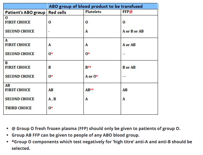
The examination aims to assess a candidate’s knowledge of:

•The basic sciences
•Clinical anaesthesia (including obstetric anaesthesia & analgesia)
•Resuscitation and emergency medicine
•Specialist anaesthesia (e.g. neuro-, cardiac, thoracic, paediatric)
•Intensive care
•Management of chronic pain
•Current literature

BASIC SCIENCES

  • anatomy, biochemistry, physiology, applied physiological measurement, pharmacology, physics and principles of measurement, STATISTICS
    CLINICAL ANESTHESIOLOGY
  • preop assessment, GA & RA , postop care NEONATAL RESUSCITATION RESUSCITATION & EMERGENCY MEDICINE
  • Basic Life Support and Advanced Life Support. Pre-hospital care. Immediate care of patients with medical or surgical emergencies, including trauma

INTENSIVE CARE AS FOLLOWS:

  • Both acute surgical and medical conditions.
  • Use of assessment and prognostic scoring systems.
  • Parenteral and enteral nutrition.
  • Biochemical disturbances such as acid base imbalance, diabetic keto-acidosis, hyperosmolar syndrome and acute poisoning.
  • Renal failure including dialysis.
  • Acute neurosurgical/neurological conditions.
  • Patients with multiple injury, burns and/or multi-organ failure.
  • Principles of ethical decision-making.

MANAGEMENT OF CHRONIC PAIN AS FOLLOWS:

  • The physiology of pain.
  • management of pain.
  • The concept of multidisciplinary care.
  • Terminal care
  • CURRENT LITERATURE

Candidates will be expected to be conversant with major topics appearing in current medical literature related to anaesthesia, pain relief and intensive care.

Whilst national and linguistic differences are recognised, some knowledge is expected on topics of international importance (e.g. new agents) even if they are not in current use in all countries.

It must he stressed that the foregoing is NOT intended either as an examination syllabus or as a comprehensive list of topics covered by the examination. It is however, a guide, which it is hoped will prove useful to candidates preparing for the diploma examination
GUIDANCE FOR CANDIDATES SITTING THE PART II EDAIC (2019)

The Part II EDAIC is an oral examination. Not all candidates are familiar with this type of examination and the following notes are intended to provide some guidance with regard both to preparation and to performance on the day.

The examination of each candidate is held in a single day during which there are four 25-minute oral examinations – (or vivas, as they are known) – two in the morning and two in the afternoon. In each of these, the candidate is examined by a pair of examiners, thereby meeting eight examiners in all. As far as possible, candidates are not examined by examiners from their own training hospital. The two morning vivas concentrate on applied basic sciences and the afternoon vivas relate to clinical topics.

Usually, but not invariably, each pair of examiners comprise one whose mother tongue is that of the language in which the candidate has chosen to be examined and the other who has a good working knowledge of the language. It is accepted that candidates may not be using their mother tongue and some allowance for linguistic difficulties is made.

In the vivas, the examiners use “Guided Questions” (GQ’s) which have been set in advance by the examination committee. Each GQ opens with a brief scenario. Ten minutes before the viva, the scenario is handed to the candidate. It is written in his/her chosen language. This gives the candidate time to collect his/her thoughts and prepare to answer questions on the topic presented. These opening questions are then followed by questions on the other topics listed in the examiner’s GQ. The first examiner asks questions for the first 12½ minutes after which a bell rings and the second examiner takes over.

Note that, whereas the Part I EDAIC basic science MCQ’s are designed to test factual recall of relevant basic science knowledge, the Part II basic science vivas are designed to test that the candidate understands the relevance of basic science knowledge applied to the practice of anaesthesia and critical care. Thus pharmacology, physiology, anatomy and relevant clinical measurement and instrumentation will always be tested. Similarly, the Part I EDAIC clinical MCQ papers are mainly concerned with testing the candidate’s factual clinical knowledge whereas the Part II clinical vivas are concerned with testing the understanding and application of that knowledge
CURRENT FORMAT OF THE EDAIC PART II EXAMINATION (2019)

The GQ’s with which the examiners are supplied list topics to be discussed with indications as to the detail required. The general format of the exam is as set out below.

MORNING

Viva 1 (Applied Basic Science)

This will start with the scenario the candidate was given 10 minutes before the start of the viva and will include applied cardiovascular and/or respiratory physiology. It will then move on to applied anatomy and physiology of other organs and systems.

Viva 2 (Applied Basic Science)

This will start with the scenario the candidate was given 10 minutes before the start of the viva and will include applied pharmacology. It will then move on to clinical measurement, applied pharmacology/physiology combined.

AFTERNOON

Viva 3 (Clinical – Critical care subject)

This will start with questions on the intensive care or emergency medicine scenario the candidate was given 10 minutes before the start of the viva. Questions on the scenario will be followed by topics such as clinical management, X-ray/CT/MRI/Ultrasound images interpretation, anaesthetic specialties and general questions.

Viva 4 (Clinical – Management of an anaesthetic problem)

This will start with questions on the anaesthetic problem scenario the candidate was given 10 minutes before the start of the viva. Questions on the scenario will be followed by questions on an internal medicine topic – possibly related to the scenario. There will also be questions on local or regional anaesthesia and some general questions.

MARKING

For each of the 20 topics of the day, each examiner can award one of three marks, which indicate respectively:

Pass ‘2’. The candidate’s performance will be deemed: fluent, able to apply knowledge, confident on core topics, thorough and able to demonstrate appropriate depth, able to correct own errors,

Borderline ‘1’. The candidate’s performance will be deemed: showing factual knowledge only (book learning with no explanation, showing poor or incomplete understanding, superficial – particularly with core topics, erratic/unstructured/disorganized, illogical but with no dangerous clinical decisions.

Fail ‘0’. The candidate’s performance will be deemed: not answering question asked despite prompting or silence, showing evidence of severe lack of topic understanding, offering multiple answers for examiner to pick, having a dangerous clinical approach

All the marks of the eight examiners (two examiners for each four sessions) will be added up to make the final score of the candidate.

To be successful, the candidate needs to obtain:
1. a score of at least 25 out of 40 in the morning sessions (Viva 1 + Viva 2)
2. a score of at least 25 out of 40 in the afternoon sessions (Viva 3 + Viva 4)
3. an overall score of at least 60 out of 80

Thus, it can be seen that, at the meeting of examiners at the end of the day, in the majority of cases there need be no further discussion of individual candidates. However, if a candidate has obtained a final score of 59, the examiners concerned would be asked to justify the mark.

Some reasons for candidates failing include:

• Inability to apply knowledge and/or basic science to clinical situations
• Inability to organise and express thoughts clearly
• Unsound judgement in decision-making and problem-solving
• Lack of knowledge and/or factual recall

In essence the examiners ask themselves the following questions:

a) Does the candidate have a good foundation of knowledge? Can the candidate apply that knowledge and understand its relevance to the practice of anaesthesia and intensive care?

b) How does the candidate approach a problem? Is the approach logical and well thought out?

c) Have alternative options been explored and understood? Is the candidate dangerous?

The Part ll examination may only be taken after the candidate has completed his/her training for specialist accreditation in their respective country. A wide general knowledge in anaesthesia, intensive care and subjects allied to anaesthesia is therefore expected.
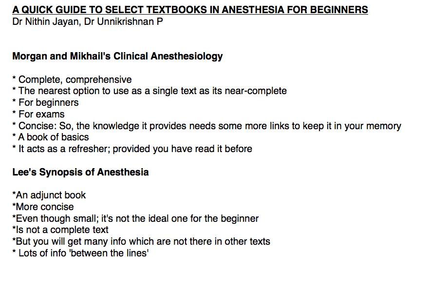
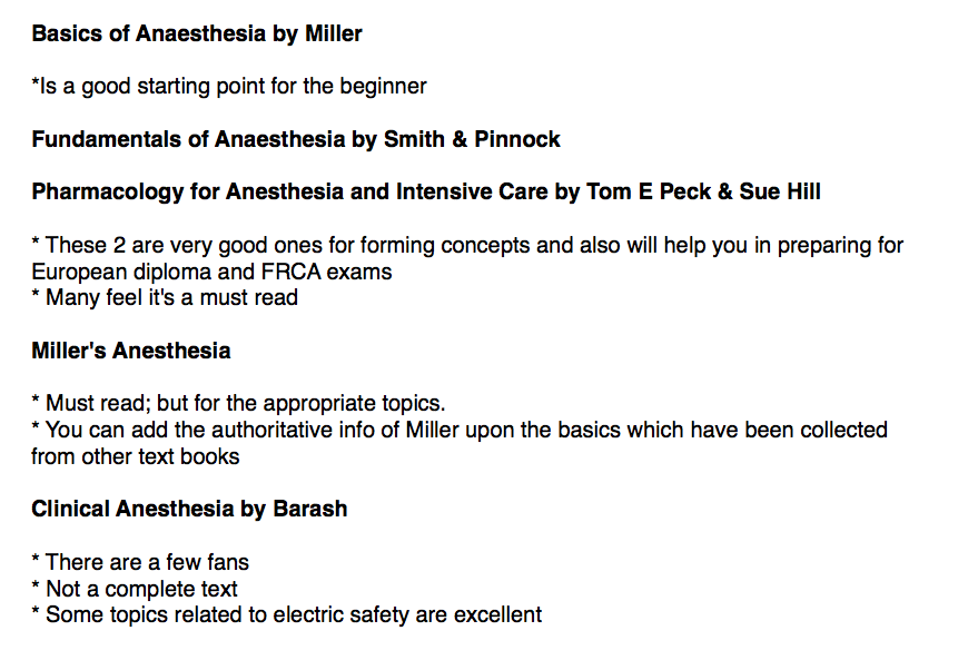
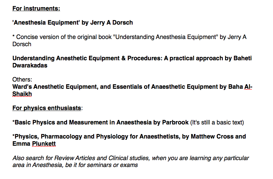
Background Reading

Which books shall I read? How much detail is required? These are common questions. There is no simple answer particularly since the EDAIC is an international exam, and the examiners and candidates come from different backgrounds. A basis for reading is the standard text book(s) of anaesthesia favoured in the candidate’s country. Familiarity with current topics from international and national journals is also be required. Access to journals may vary in different departments but the Internet now provides a wealth of new opportunities. In addition, a recommended reading is also at your disposal.

THE FOLLOWING ADDITIONAL POINTS MAY BE OF ASSISTANCE

Applied Basic Science Vivas

Physiology

It is obvious that the physiology of the cardiovascular and respiratory systems will be examined in some detail. A good knowledge of neuro, renal and hepatic physiology as applied to anaesthesia and intensive care will also be expected. Other areas relevant to anaesthesia will also be covered but great detail is not expected.

Pharmacology

The principles of pharmacokinetics and pharmacodynamics will be examined in some detail. An intimate knowledge of the pharmacology and toxicology of drugs used in anaesthesia is expected as well as many of the drugs in common use in intensive care. An informed anaesthetist who reads journals must have some understanding of research protocols and the relevance of statistical methods employed, in order to judge the value of articles.

Applied Anatomy

It is expected that anaesthetists will know the essential anatomy of areas into which they may insert needles cannulae and endotracheal and endo-bronchial tubes. Applied anatomy of the heart and lung is also examined.

Physics and Clinical Measurement

Anaesthetists monitor and measure numerous clinical parameters and take action on the information displayed. It is expected therefore that they should understand the principle of action, limitations, accuracy, and sources of error in these monitors. Some of the basic physics of gases and vapours, and principles of electrical safety are essential knowledge for the informed anaesthetist. The principle of action and causes of failure in anaesthetic machines and ventilators is also essential knowledge.

Clinical Anaesthesia & Intensive Care Vivas

Clinical Anaesthesia

As candidates will have completed their training to the standard required for specialist registration they should have experience in all types of anaesthesia and intensive care. These vivas will include questions on both general, regional and special anaesthetic techniques as applied to neuro-, cardiac and paediatric surgery, obstetric anaesthesia and the management of acute and chronic pain.

The examiners do not have direct experience of how the candidate would deal with an anaesthetic problem. They therefore have to make a judgement based upon the candidate’s performance in the oral exam. The examiner cannot assume the candidate would have carried out a procedure or checked a clinical or electronic monitor. The candidate must mention it.

Clinical scenario

An example of the clinical scenario given in advance to a candidate would be as follows: A 67-year-old man weighing 100kg, 1.67m in height is scheduled for an elective repair of a 10cm abdominal aortic aneurysm. He had myocardial infarction 6 months previously and has been a non-insulin dependent diabetic for over 10 years. Discuss your anaesthetic management of this case.

The initial discussion on this sort of opening scenario will reveal much about the candidate’s approach to the problem and an awareness of the potential dangers. Remember that the anaesthetic management starts in the ward!

Definition of problems: Clearly, the primary problem is the presenting aneurysm and its repair. What will it involve?

Secondly the patient is obese and has, as yet unquantified, cardiovascular problems and diabetes.

This would lead to a full medical history with emphasis on the above with appropriate examination and investigation of potential complications. The anaesthetic management would involve choice of technique, appropriate monitoring, management of complications and post-operative pain relief.

A candidate who presents a logical well structured answer, explaining the reasons behind the proposed course of action, is more likely to find that the examiner says very little and does not have to interject continually. It cannot be emphasised enough that practice in presentation is essential and candidates should practice this skill with their trainers or fellow trainees. This is even more important for candidates not using their mother tongue

This topic alone, could take up more than the allotted time and so examiners may suddenly curtail discussion on a given subject and move on to something else. This is a necessary part of the examination process and does not indicate displeasure with the answers given.

Candidates should appreciate that the intention of the examiners is to enter into a dialogue with them regarding whatever topic is under discussion. The intention is not simply to find the candidate’s areas of ignorance although, inevitably, these may become apparent – if they exist. Bearing this in mind, the candidate should try to discuss the topic knowledgeably and should not be afraid to say when the topic is completely outside his/her experience. The EDAIC being an international exam and not a collection of national exams, means inevitably, that a wide range of views will be held both by the candidates and examiners.

It is assumed that candidates have been trained in standard mainstream anaesthetic techniques. They would be wise therefore to base their answers on methods with which they are familiar and would be normal in their institution, rather than straying into unfamiliar territory in the mistaken belief that this might be the answer the examiners require. Examiners will sometimes query an answer to see whether the candidate is confident in their answer or can be swayed from their course of action. There will often be no right or wrong answer to a question and examiners will accept an answer or opinion that is based on sound evidence and justifies the proposed course of action

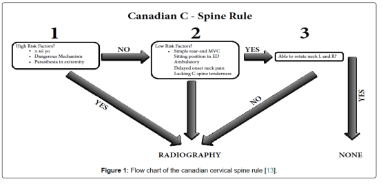
SYSTEMATIC REVIEW OF

Images

Candidates are expected to have a systematic and logical approach to reading Images and should be able to describe their system to the examiner. A typical system would be:

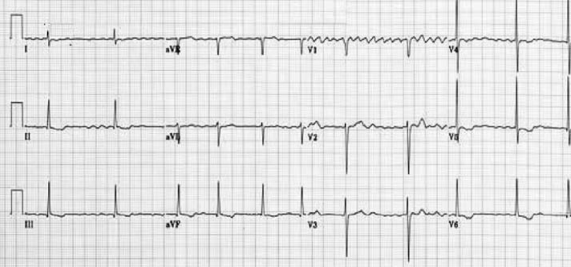
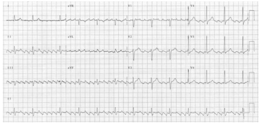
For example, for X-rays:

Markings: Look at writing on the film: name/age of the patient and projection of the radiograph.

Film Quality: Penetration, rotation & inspiration (on a chest film).

Review Areas: Lungs, diaphragm, pleura, upper abdomen, heart & mediastinum, bones of thoracic cage & soft tissues.

Artifacts: Note the presence of any equipment placed in the chest by anaesthetists or surgeons!

Recognition of Critical Incidents and taking prompt and appropriate action

One common cause for failure in the exam is a haphazard approach to dealing with critical situations that are posed and not following Advanced Life Support protocols. Airway, Breathing and Circulation should be the foundation of all resuscitation.

Diagrams & Graphs

Use of diagrams, graphs and other material to present answers. Pencils and paper are provided at all times during the Part II vivas. Candidates can use them to advantage in making presentations and explaining points. A typical scenario given in advance in the applied basic science exam might be: Discuss the factors that influence carriage of oxygen in the blood. A diagram of the various oxy-haemoglobin dissociation curves with some relevant values would create a good impression at the commencement of the exam and help the candidate settle into a structured answer. In pharmacology, the value of diagrams and graphs in explaining the principles of pharmacodynamics or pharmacokinetics is obvious .

N.B. For EDAIC 1
1-2 Questions from statistics will come in Part 1 Paper A

Statistics: Basic principles of data handling, probability theory, population distribution and the application of both parametric and non-parametric tests of significance.

GENERAL ADVICE ON PRACTISING FOR VIVAS (Basically for FRCA; but may be useful for EDAIC too)


We found the following techniques extremely valuable in the run-up to the vivas:

1.Group revision
2.Frequent practice
3.Practise categorising
4.Card system

  • Group revision: It is extremely useful to team up with some friends or colleagues regularly in the weeks before the viva and practise talking about anaesthetic topics. Practising with friends has several advantages: Seeing your friends on a regular basis will help keep you sane. This is better than locking yourself in a small room with a pile of books and trying to learn the coagulation cascade for the fifth time since qualification! Your morale will remain in better shape than if you were revising on your own because you will be able to encourage each other. You will also be more aware of the progress you are making. As a group, you can pool your resources in terms of reference books and previous questions. During the working day, one of you may have had a practice viva with a consultant who asked an awkward question or a common question asked in a different way. You can then discuss with your friends how they would have answered it. Different people revise in different ways and, consequently, will have their own way of talking about a subject. This means that others in the group will benefit from listening to the practice viva. They may have a particular piece of knowledge that really helps an answer gel together or they may use a particular turn-of-phrase that succinctly deals with a potential minefield. You can practise phrasing your answers in a particular way in the knowledge that, if it all falls apart halfway through, it won’t matter and you can have another go. This is less easy to do in front of consultants who might write your reference! By being ‘the examiner’, you will gain insight into the pitfalls of the viva process. You can usually see someone digging a hole for themselves a mile off!
  • Frequent practice: Repetition of clinical scenarios. During your revision, you will find the same clinical situations coming up time and time again (as in the exam). Over the years, anaesthetic techniques may change but new techniques are all aimed at trying to solve particular clinical problems, for example, the fibre-optic scope to help with the difficult airway or new drugs that provide more cardiovascular stability. However, the problems remain the same! Patients will still present with difficult airways, ischaemic heart disease, COAD, obesity, hypertension, etc. The more you practise, the more often you will find yourself repeating the problems each of these scenarios presents and thus the more confident and slick you will become at delivering the salient points. There are obviously a few exceptions, e.g. MRI scanners and laser surgery, where the advancement of technology has presented new challenges to the anaesthetist. These situations are in the minority and as long as you are aware of them and the associated anaesthetic problems, you should be well-equipped to deal with questions on them in the exam.
  • The clinical scenarios break down into a few categories:
  • Medical conditions that have anaesthetic implications, e.g. Aortic stenosis, Diabetes, Hyperthyroidism.
  • Surgical procedures that have anaesthetic implications, e.g. Oesophagectomy, CABG, Pneumonectomy.
  • Anaesthetic emergencies/difficult situations, e.g. Anaphylaxis, Malignant hyperthermia, Failed intubation. Paediatric cases. These represent a limited range of cases the examiners are likely to ask you about, e.g. Upper airway obstruction, Pyloric stenosis, Bleeding tonsil.
  • Having repeatedly practised these clinical scenarios, you will soon realise that the problems of anaesthetising an obese patient with diabetes, ischaemic heart disease, porphyria and myasthenia for an abdominal aortic aneurysm repair (!) can be broken down into the problems that the respective conditions present to the anaesthetist, plus the problems of the specific operation. You may then approach what seems to be a nightmare question with a degree of confidence and structure.
  • Phrasing: It cannot be over-emphasized that frequent practice will improve your viva technique. As already mentioned, some topics crop up again and again in different situations, such as part of a long case or even a complete short case (e.g. obesity, anaesthesia for the elderly or the difficult airway). With regular practice, you will soon develop your own ‘patter’ to help you deal with these common clinical scenarios. These can then be adopted at opportune moments to buy yourself easy marks whilst actually giving you time to gather your thoughts
  • Practise categorising: Putting order to your answers demonstrates to the examiners that you conduct your clinical practice in a systematic and safe way. If you do not mention the most important points first (e.g. airway problems in a patient presenting with a goitre), then this may suggest to the examiners that you are disorganized. An ‘ABC’ (order of priority) approach to many of the questions may be helpful. For example, in obese patients, managing the airway has a higher priority than difficulty with cannulation. It is often a good idea to use your opening sentence to tell the examiners how you are going to categorize your answer.

Example 1:

‘Tell me about the anaesthetic implications of rheumatoid arthritis’.

‘Patients with rheumatoid arthritis may have a difficult airway and secondary respiratory and cardiovascular pathology. They are frequently anaemic, taking immunosuppressant drugs and the severe joint pathology leads to problems with positioning’.

Example 2:

‘What are the important considerations when anaesthetising a patient for a
pneumonectomy’?

‘These may be divided into three broad areas: the pre-operative assessment of fitness for pneumonectomy and optimisation, the conduct of anaesthesia with particular reference to one-lung anaesthesia, positioning, intra-operative monitoring and fluid balance and finally post operative care’.

  • Card system: We formatted postcards to summarise the main problems associated with different anaesthetic situations. These proved to be a good starting point for viva practice and a quick source of reference. They also encouraged us to deliver the first few points in a punchy manner.

For example:
‘What problems do you anticipate with anaesthetising a patient with Down’s syndrome’?

‘These patients present the following problems for the anaesthetist. They may have a difficult airway, an unstable neck, cardiac abnormalities, mental retardation, epilepsy and a high incidence of hepatitis B infection’.

VIVA TECHNIQUE

  • 1.Think first: Don’t panic. If you are unlucky enough to be asked a question about an obscure subject such as lithium therapy (as two of us were in our science viva), remember the examiners have only just seen the questions as well. It may also be of some comfort to know that there will be at least ten other candidates being asked the same question at the same time. Keep things simple at first and think about how you are going to structure your answer. Categorising your answer may allow you to deliver more information about the topic than you thought you knew. Conversely, do not dwell on what you do not know, e.g. the pH and dose!

Example: ‘Tell me about lithium’
Think . . . ‘What is it used for’?
Say . . . ‘Lithium is a drug used in the treatment of mania and the prophylaxis of manic depression’.

Think . . . ‘What is the presentation and dose? . . . I don’t know the dose’.
Say . . . ‘It is presented in tablet form’.

Think . . . ‘What is its mode of action? . . . I have no idea but I know it is an antipsychotic’!
Say . . . ‘Its main action is as an antipsychotic’.

Think . . . ‘Why are they asking me this question? What is the relevance to anaesthetic practice’?
Say . . . ‘It has a narrow therapeutic range and therefore toxicity must be looked for. Side effects may include nausea, vomiting, convulsions, arrhythmias and diabetes insipidus with hypernatraemia’.

A similar approach can be used for the clinical viva.

  • The opening sentence : This will set the tone of the viva. If the first words to come from your mouth are poorly structured, ill thought-out or just plain rubbish, then you are likely to annoy the examiners and will face an uphill struggle. If, on the other hand, your first sentence is coherent, succinct and structured, then you will be half-way there. With a bit of luck, the examiners will sit back, breathe a sigh of relief (because it has been a very long day for them) and allow you to demonstrate your obvious knowledge of the subject in hand!

For example:

‘What are the problems associated with anaesthesia for thyroid disease’?
‘Anaesthesia for patients with thyroid disease has implications in the pre-, intra- and post-operative periods’.
You are then able to expand in a logical way from here.
‘Pre-operatively, assessment of the airway and control of the functional activity of the gland is essential . . . ’

  • Categorise or die!

Remember this lends structure to your answer and gives the examiners the impression you are about to talk about the subject with authority. If you categorise your answer well enough, they may actually stop you and move onto something else.

  • Opening question

You will be asked to summarise the case so prepare your opening sentence
beforehand.

For example:
You may be asked to summarise the scenario of a 75-year-old man with chronic obstructive pulmonary disease who is scheduled to undergo an elective cardio-oesophagectomy the following day.

‘Would you like to summarise the case’?

One possible answer may begin:

‘This is an elderly gentleman with complex medical problems who is scheduled for a cardio oesophagectomy. He has evidence of chronic obstructive pulmonary disease, ischaemic heart disease and diabetes. There will be substantial strain on his cardio-respiratory system. This operation is a major procedure that involves considerable fluid shifts, a potential for large blood loss and requires careful attention to analgesia. These are the main issues that I would concentrate on in my pre-operative assessment’.

Even though a cardio-oesophagectomy involves other considerations (e.g. double-lumen tube / one-lung ventilation) it can be seen that this opening sentence could be adapted to suit other clinical scenarios such as:

Pneumonectomy
Laparotomy
CABG / valve replacement
Cystectomy
Open prostatectomy

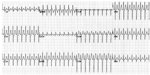
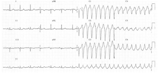
  • Analyse all the investigations

You will be asked for your opinion on the ECG, chest X-ray, blood results, etc., so make sure you have decided on the abnormalities and the most likely causes for them in the 10 minutes you have to view the data. Try to make your answers punchy and authoritative.

For example, ‘The ECG shows sinus rhythm with a rate of 80 and an old inferior infarct’ is better than going through the ECG in a painstaking ‘The rate is . . . the rhythm is . . . the axis is . . . ’

Don’t waste valuable time waffling on about the normal-looking bones on a chest X-ray if there is a barn-door left lower lobe collapse. This does not necessarily imply you are not thorough, providing you demonstrate that you have looked for and excluded other abnormalities.

  • Anaesthetic technique

You will usually be asked how you would anaesthetise the patient in the long case. There will often not be a right or wrong answer, but you should try to decide on your technique and be able to justify it. The examiners may only be looking for the principles of anaesthesia for a particular condition such as aortic stenosis, although this is probably more likely in the short cases.

For example:
‘You are asked to provide an anaesthetic for a 77-year-old lady who needs a hemi-arthroplasty for a fractured neck of femur. She had a myocardial infarction 3 months ago and has evidence of heart failure’.

You should be able to summarise the principles involved and choose an anaesthetic technique appropriate to the problems presented. You could, for example, give this patient a general anaesthetic with invasive monitoring , you could use TIVA with remifentanil or a neuroaxial block.
All of these techniques could be justified, but to simply say that you would use propofol, fentanyl and a laryngeal mask without saying why, may be asking for trouble!

In some circumstances it may be the options for management rather than a specific technique that is required. You may find it appropriate to list the options for analgesia in a patient having a pneumonectomy, for example, and then say why you would use one technique over the others.
You should try to address the anaesthetic technique for the long case BEFORE you face the examiners. You will not look very credible if you have had 10 minutes to decide on this and have not reached some kind of conclusion. Overall, most candidates felt that the examiners were pleasant and generally helpful. If you are getting sidetracked they will probably give you a hint so you do not waste time talking about something for which there are no allocated marks. If they do give you a hint, take it!

Good luck.