VIVA SCENE: PARACETAMOL Vs MORPHINE CENTRAL MOA

PARACETAMOL:

Central action via COX 3 inhibition which is associated with decreased brain PGE2 levels. It also modulates endogenous cannabinoid system

MORPHINE:

Morphine work by stimulating presynaptic Gi-protein-coupled MOP and KOP opioid receptors. Binding of the ligand causes the following events:

> Closure of voltage-gated Ca2+ channels

> Decreased cAMP production

> Stimulation of K+ efflux from the cell

> Hyperpolarisation of the cell membrane.

> This leads to decreased excitability of the cell and therefore decreased neurotransmitter release and pain transmission

VIVA SCENE: Ideal Inhalational Agent

Screen Shot 2019-09-05 at 7.20.43 pm

VIVA SCENE: THROMBOELASTOGRAPHY (TEG) AND OTHER QUESTIONS

TEG is a relatively new modality for monitoring coagulation which is very useful during management of trauma and also in the perioperative scenario..

BASIS:

  • The 2 main components of the TEG machine are a cup and a pin. Whole blood is mixed with the activating agent kaolin as well as calcium. The cup then oscillates around the pin slowly, at a rate of 6 times per minute, to mimic natural blood flow in vivo and activate the clotting cascade. As the clot forms, the torque between the cup and pin is transduced and measured, creating a curve. As the clot breaks down and torque decreases, the tracing converges to represent this. 
  • The different parameters of the curve are then measured to assess current coagulation status.
  • Of the 4 types of TEG assays available, the most common is the rapid TEG. The use of an activator in rapid TEG standardizes the TEG test and speeds up the rate at which clotting takes place, thus making results available more quickly.

INTERPRETATION

Screen Shot 2019-09-04 at 3.20.39 pm

Screen Shot 2019-09-04 at 3.04.41 pm

  • R(sec): The first measurement of note is the reaction time (R time). This is the time interval from the start of the test to the initial detection of the clot. Normal R values range between 7.5 and 15 minutes. A prolonged R time may indicate hemodilution or clotting factor deficiencies. The treatment for prolonged R time is to administer FFP as it contains all factors of the coagulation cascade, without further coagulant hemodilution A shortening of R time (< 3 minutes) occurs in hypercoagulable states. Examples would be patients with early disseminated intravascular coagulation (DIC) or septicemia. In these situations, free thrombin is released into the circulating blood, triggering the clotting mechanisms but the patient later begins to bleed because of exhaustion of clotting factors.

  • K (sec) and Angle α (°): The clot strength is measured by these 2 variables in TEG. The K value measures the interval between the R time and the time when the clot reaches 20 mm. Normal K values range between 3 and 6 minutes. Prolongation of the K value with normal platelet count  indicates inadequate amounts of fibrinogen to form fibrin. The treatment for prolonged K value is therefore to administer fibrinogen/cryoprecipitate. The α angle measures a line tangent to the slope of the curve during clot formation.The alpha angle represents the thrombin burst and conversion of fibrinogen to fibrin. Normal α value is between 45° and 55°. A longer K value causes a shallow or more acute angle (<45°), while a shorter K value causes a steeper α angle (>45 °). An angle α <45° suggests a less vigorous association of fibrin with platelets. In this case, treatment begins much higher on the coagulation cascade, with the replacement of both fibrinogen and factor VIII. Thus, these patients can be treated with the administration of cryoprecipitate. Shortening of the K-value indicates a very quick formation of clot, potentially due to hypercoagulability or inappropriate consumption of coagulation factors. A shortened K value also corresponds to a steeper α (>45°). The treatment for shortened K and steeper α is anticoagulation therapy

  • MA (mm): Maximum amplitude is a measurement of maximum clot strength and provides information on both fibrinogen and platelet function. As the clot develops and increases in tensile strength due to platelet activation and binding to fibrin, the tracing increases it’s MA or appears to widen. Normal values are between 50–60 mm. 80% of the MA is derived from platelet function whereas the remaining 20% is derived from fibrin. A low MA value is indicative of low clot strength, which can be caused by decreased fibrinogen levels, low platelet counts, or decreased platelet function. (i) Paired with a prolongation of K value, this could be a sign of the need for cryoprecipitate. (ii) Administration of platelets may be avoided when a low platelet count is combined with a normal MA value (=platelet function is normal) (iii) Treatment with platelets may be indicated for patients with a low MA value (=low platelet function) and normal platelet count. (iv) High MA will occur in the setting of hyperactivity of platelets, and MA above 75 mm indicates a prothrombotic state. In this case, treating with an anticoagulant would be helpful
  • Shear Elastic Modulus Strength, G value or G: is a measure of clot strength or clot firmness, and is calculated based on the amplitude value (A) until the maximum amplitude (MA) is reached. It is the single most important value of the entire assay because it represents the overall function or effectiveness of the clot. Normal G values are between 5.3 and 12.4 dynes/cm2. A G value >10 dynes/cm2 indicates increased risk of thrombosis. Treatment for high G is accomplished by the use of platelet inhibitors such as Clopidogrel or Aspirin. Aspirin is usually not preferred because it inhibits platelet adherence rather than platelet aggregation. A G <5 dynes/cm2 places a patient at increased risk of hemorrhage
  • As time progresses during the TEG assay, the tracing will remain at maximal amplitude for a period of time, after which clot lysis begins. Normally, lysis continues for a period of up to 15 minutes. A computerized algorithm automatically estimates the percentage of lysis occurring over time. This is called the Estimated Percentage of Lysis or EPL. After 30 minutes, EPL becomes EPL30 or succinctly LY30 (i.e. percentage of lysis at 30 minutes). Both the EPL and LY30 are measurements of excessive fibrinolysis since they measure the percentage decrease in amplitude after MA. An EPL between 7.5 and 15%, when accompanied by a very high G, reflects a hyperfibrinolytic and hypercoagulable state typical of patients with early DIC. A very high EPL or LY30 (>20%) may indicate the need for antifibrinolytic therapy, such as the use of transexamic acid or aminocaproic acid. LY30 is also useful for patients undergoing thrombolytic drug therapy. This can be observed by rapid curve convergence.

Ref: Thromboelastography: Clinical Application, Interpretation, and Transfusion Management, Shawn Collins et al AANA Journal Course, 2016

HOW DO WE TEST CLOTTING?

  • By doing tests like aPTT, PT & INR, Platelet Count, ACT, Bleeding Time, fibrinogen and factor levels, TEG etc
  • The aPTT and INR use different reagents to measure the time to form a clot in vitro after platelet-poor plasma from blood collected in a calcium chelating tube, is recalcified
  • The aPTT is prolonged with the deficiency of factors of the intrinsic pathway: Fs 8,9,11,12. Also the factors involved in the common pathway (Fs 1,2,10)e.g. Heparin therapy, DIC, liver disease
  • The INR is prolonged especially with deficiency of F 7; but also with deficiency of Fs 1,2,5,10 e.g.  warfarin therapy, vitamin K deficiency, DIC, liver disease
  • N.B Warfarin inhibits the gamma carboxylation of vitamin K dependent factors 2,7,9,10
  • BLEEDING TIME :Duke’s method: Sterilize the finger tip using rectified spirit and allow to dry. Make a sufficiently deep prick using a sterile lancet, so that blood comes out freely without squeezing. Note the time (start the stop-watch) when bleeding starts. Mop the blood by touching the finger tip with a filter paper. This is repeated every 15 seconds, each time using a fresh portion of the filter paper, till bleeding stops. Note the time (stop the stop-watch). Normal value is upto 4 minutes. 
  • CLOTTING TIME: Capillary tube method: (Wright’s method). Under sterile precautions make a sufficiently deep prick in the finger tip. Note the time when bleeding starts (start the stop watch). Touch the blood drop at the finger tip using one end of the capillary tube kept tilted downwards. The tube gets easily filled by capillary action. After about two minutes start snapping off small lengths of the tube, at intervals of 15 seconds, each time noting whether the fibrin thread is formed between the snapped ends. Note the time (stop the stop watch) when the fibrin thread is first seen. Clotting time is the interval between the moment when bleeding starts and the moment when the fibrin thread is first seen.

    Normal value is 3 to 10 minutes.

  • Bleeding time depends on the integrity of platelets and vessel walls, whereas clotting time depends on the availability of coagulation factors

VIVA SCENE: CARBON MONOXIDE (CO) POISONING

AETIOLOGY: Carbon monoxide is produced by incomplete combustion and is found in car exhaust, faulty heaters, fires and in industrial settings. Carboxyhaemoglobin (COHb) concentrations in cigarette smokers range as high as 10%.

MECHANISM: Binds to Hb with 210 times affinity than O2: so reduce the O2 carrying capacity of blood. Also disrupts oxidative metabolism, binds to myoglobin and cytochrome oxidases, causes lipid peroxidation. Final result is tissue hypoxia. Severity depends on the duration of exposure, CO levels and patients pre-event health status: pre-existing cerebral disease, cardiac failure, hypovolemia and anemia increase toxicity

DIFFERENTIAL DIAGNOSIS: Cyanide poisoning ( suspected when CNS effects are out of proportion with COHb concentrations and if there is a marked lactic acidosis)

Screen Shot 2019-09-04 at 11.31.29 am

EVALUATION & MANAGEMENT:

  • ABC approach
  • Secure the airway; if GCS<8, consider intubation
  • Stabilize respiration:  consider mechanical ventilation or CPAP
  • Get intravenous access
  • Send samples for estimation of Hb(?anemia), electrolytes (dyselectrolytemias worsen the cardiac toxicity), COHb levels (to confirm diagnosis; useless in prognosis), blood sugar, ABG and cardiac enzymes. Take an ECG.
  • Metabolic acidosis due to lactate give a clue to the extend of ischemia. Net effect of metabolic acidosis may be beneficial on O2 delivery; but treated if pH<7
  • Patient discouraged from activity
  • 100% O2 reduces the half life of COHb from 4 hours (in ambient air) to 40 minutes. 4 to 6 h of 100% normobaric oxygen will remove over 90% of the carbon monoxide. Oxygen toxicity is unlikely with less than 24 h treatment
  • When immediately available, hyperbaric oxygen (HBO) should be considered with serious CO poisoning. Oxygen at 2–3 atmospheres will further reduce the half-life of COHb to about 20 min but, more importantly, it causes very rapid reversal of tissue hypoxia due to oxygenation of tissue from oxygen dissolved in the plasma. Some clinicians implement it based on the presence of any of the following: history of loss of consciousness, abnormal neuropsychiatric testing or neurological signs, pregnancy. COMPLICATIONS OF HBO: decompression sickness, rupture of tympanic membranes, damaged sinuses, oxygen toxicity

  • CO PRODUCTION WITH SODALIME USE: Occurs when inhalational agents with CHF2 moiety such as desflurane, enflurane, and isoflurane are used with desiccated soda lime granules that was left unused for a long time. Can be significant in smokers especially when very low flows are used. Factors increasing the production of CO include° Type of inhaled anaesthetic agent (magnitude of CO production from greatest to least is desflurane > enflurane > isoflurane > sevoflurane)° High absorbent dryness ° Type of absorbent (at a given water content, baralyme produces more CO than soda lime)° Increased temperature° Higher anaesthetic concentration

VIVA AID: MAO INHIBITORS AND OPIOIDS


  • Some opioid analgesics are associated with a risk of serotonin syndrome in combination with MAOIs due to their serotonergic properties. Other combinations may result in opioid toxicity due to CYP450 enzyme inhibition by the MAOI.
  • Given the widespread availability of several suitable alternative drugs, the combination of dextromethorphan, methadone, pethidine, tramadol, fentanyl or tapentadol with an MAOI should usually be avoided, including in the 14 day period following the withdrawal of an irreversible MAOI.
  • Morphine, codeine, oxycodone and buprenorphine are alternative opioids for patients receiving MAOIs, though starting at a low dose and titrating cautiously against clinical response is advised. Blood pressure and the signs and symptoms of CNS and respiratory depression should be monitored closely.
  • The MAOI/pethidine interaction has two distinct forms: an excitatory and a depressive form. Pethidine must never be used in the presence of MAOIs because of the risk of a fatal excitatory interaction. Morphine does not cause this excitatory interaction, and is the drug of choice provided an allowance is made for possible potentiation of the depressive narcotic effect.


VIVA SCENE: C-SPINE X-RAY

WHETHER TO DO C SPINE IMAGING IN TBI; CRITERIAS:

1. Screen Shot 2019-08-11 at 9.23.39 pm

2. Under the NEXUS guidelines, when an acute blunt force injury is present, a cervical spine is deemed to not need radiological imaging if all the following criteria are met:

  • There is no posterior midline cervical tenderness
  • There is no evidence of intoxication
  • The patient is alert and oriented to person, place, time, and event
  • There is no focal neurological deficit (see focal neurological signs)
  • There are no painful distracting injuries (e.g., long bone fracture)

Screen Shot 2019-08-26 at 11.12.53 am

Lateral C-Spine Radiograph. (AABCDs)

A—Adequacy: An adequate film should include all seven cervical vertebrae and C7/T1 junction with optimum density so that the soft tissue shadow is visible clearly.

A—Alignment

a. Atlanto occipital alignment: The anterior and posterior margins of the foramen magnum should line up with the dens and the C1 spinolaminar line.

b. Vertebral alignment

Look for the following four lines (any incongruity= should be considered as evidence of ligamentous injury or occult fracture)

1. Anterior vertebral line—joining the anterior margin of vertebral bodies

2. Posterior vertebral line—joining the posterior margin of vertebral bodies

3. Spinolaminar line—joining the posterior margin of spinal canal

4. Spinous process/ Interspinous line—joining the tips of the spinous processes

B Bony Landmark: vertebral bodies, pedicles, laminae, and the facet joints are inspected

C Cartilagenous space: Predental space or the Atlanto-Dental Interval (ADI): which is the distance from dens to the body of C1. ADI should be < 3 mm in adults and < 5 mm in children. An increase in ADI depicts a fracture of the odontoid process or disruption of the transverse ligament

D—Disc space

Disc spaces should be roughly equal in height and symmetrical.

Loss of disc height can happen in degenerative diseases.

S—soft tissue

Prevertebral soft tissue space thickness can help in the diagnosis of retropharyngeal haemorrhage, which can be secondary to vertebral fractures. Maximum allowable distances :

Nasopharyngeal space (C1): 10 mm

Retropharyngeal space (C2–C4): 5–7 mm

Retrotracheal space (C5–C7): 14 mm in children and 22 mm in adults

VIVA SCENE IMAGING: CHEST X-RAY (CXR)

  • Helpful MNEMONICS: ‘PRIP ABCDEFGHI’. Also in inspiratory films the level of the diaphragm is at the level of ribs 5/6 anteriorly and 8/10 posteriorly. (AR6PR10). 
  • Right heart border is formed predominantly by the right atrium along with the lower part of SVC, whilst the left border is formed by the aortic arch, pulmonary artery, left atrium, and ventricle. The right and left ventricle forms the inferior border. The right diaphragm is normally higher than the left due to the liver.
  • Areas where pathology is commonly missed: Apices (including behind the 1st rib and clavicle)—small pneumothoraces and masses. Hila—masses and lymph nodes; left hilum is 1–2 cm higher than right. Behind the heart—left lower lobar collapse and hiatus hernia. Below the diaphragm—free gas. Soft tissues—breast shadow or absence (look for lung and bone metastasis)
  • This is CXR of Mr Roy (Patient identity) done on 14/08/2019 (date of study)
  • Its an AP film (Projection)
  • Non rotated (Rotation)
  • Expiratory film (Inspiratory/Expiratory)
  • Adequately penetrated (Penetrated)
  • Trachea is central, no foreign bodies or other abnormalities (A– Airway)
  • On both sides, the bones and soft tissues appear normal; ribs, sternum, scapula, clavicle, spine, and humerus has no deposits or fractures (B-Bones and soft tissues)
  • The cardiac silhoutte is normal (C-Cardiac silhoutte)
  • There is no free air under the diaphragm. Bilateral costo- and cardio-phrenic angles are clear (D– Diaphragm and angles)
  • No effusions. Pleura is not visible (E-Effusion)
  • Both sides, the lung fields appear normal (F-Fields = lung fields)
  • Gastric bubble visible at left upper abdomen (G=Gastric bubble)
  • The hila and the mediastenum appears to be normal and not displaced. The CT ratio is less than 50% (H-Hila and mediastenum)
  • Endotracheal tube tip is seen 5 cms above the carina or T4/T5 interspace. NG tube passed down midline, past level of diaphragm, and deviates to left with tip seen in stomach. ECG leads and electrodes are noted (I=Insertions, Interventions: tubes and lines, chest drains, pacemakers, and metallic valves and artefacts)
Screen Shot 2019-08-25 at 5.10.08 pm
  • If an intrathoracic opacity is in anatomical contact with the heart border, then the opacity will obscure that border. If an intrathoracic opacity is in the posterior pleural cavity so not in direct anatomical contact with the heart border, this causes an overlap but not an obliteration of that border (Silhoutte sign)
  • When the alveoli no longer contain air and opacify, the air-filled bronchi passing through the alveoli may be visible as branching linear lucencies. (Air bronchogram)
  • Linear opacities measuring 1–6 cm extending from periphery to the hila caused by distension of anastomotic channels between peripheral and central lymphatics.(Kerley A lines)
  • Short horizontal lines, due to oedema of the interlobular septae, situated perpendicularly to the pleural surface at the lung base (Kerley B lines)
  • Reticular opacities at the lung base (Kerley C lines)
  • The mediastinal masses can be identified on the PA views but lateral films and/or CT scans are required to confirm further diagnosis
  • Anterior mediastenum: Anterior to trachea, Middle mediastenum: Anterior to heart, Posterior mediastenum: Posterior to heart
  • ANTERIOR MEDIASTENAL MASS: (4 T’s) Thymic tumour -Teratoma- Thyroid –  Terrible lymph nodes (LN) Ascending aortic aneurysm, MIDDLE MEDIASTENAL MASS (BAL) Bronchogenic cyst – Arch aneurysm- LNs, POSTERIOR MEDIASTENAL MASS: as expected it can be from Spine/Neurogenic; also hiatus hernia
  • Hilar mass: Most commonly TB/Sarcoidosis; also lymphoma
  • Collapse: Triangular opacity, Crowding of ribs, Mediastenal displacement, hyperinflation of other lobes
  • Consolidation: confluent ill defined opacity, bat wing distribution, no volume loss, air bronchogram
  • Effusion: Erect: obliteration of the costo- and cardio-phrenic angles and opacity with meniscus. Supine: graded haze giving a ground glass opacity
  • Emphysema: Decreased lung markings, flat diaphragm and hyperexpanded lung, Presence of bullae and peribronchial thickening, signs of cor pulmonale, right ventricular enlargement, pulmonary hypertension—enlargement of the central pulmonary arteries with oligaemic peripheral lung fields

VIVA SCENE : NEONATE /OLDER CHILD Vs ADULT

CVS:

  • Average HR is 120-180 bpm and SBP 50-90 mm of Hg at birth
  • Oxygen consumption is twice that of an adult 7 ml/kg/min. But because of a fixed stroke volume, increasing the HR is the only way to increase CO. Also neonatal myocardium consists of more non-contractile connective tissue.
  • Parasympathetic system is predominant; bradycardia can happen in response to hypoxia or tracheal suction. Asystole is the most common form of cardiac arrest and ventricular fibrillation is uncommon
  • Circulating volume approx 90 ml/kg @ birth (300-400 ml in an average neonate). Bleeding contributes proportionately a greater loss of total volume in a neonate, compared to an adult. Right ventricular mass equal to left ventricular mass until 6 months of age, resulting in right axis deviation on the ECG.
  • Transitional circulation: Before birth the SVR is low due to the low resistance placental circulation whereas the pulmonary resistance is high. At birth the SVR rapidly increases after clamping of the umbilical cord and PVR decreases following functional closure of foramen ovale (due to increased venous return to left atrium from pulmonary arteries) and ductus arteriosus (in response to increased PaO2 in blood). But this can be reversed back to the foetal state due to stimuli including hypoxia, heypecarbia and acidosis, leading to a perpetuating cycle of worsening hypoxia. This is known as transitional circulation or prsistent foetal circulation.

AIRWAY/ RESPIRATORY SYSTEM

  • Relatively larger head, short neck, large tongue and narrow nasal passages.
  • High anterior larynx (level C2/3 compared to C5/6 in the adult).
  • Large U-shaped floppy epiglottis.
  • Narrowest point of the larynx is at the level of the cricoid cartilage (in adults it is at the laryngeal inlet).
  • Trauma to the small airway can easily lead to oedema and airway obstruction. 1 mm oedema can narrow an infant’s airway by 60% (resistance ∝1/radius).
  • Equal angles of mainstem bronchi (in adults the right main bronchus is more vertical).
  • A compliant chestwall: FRC is less as the elastic recoil pulls the compliant chest wall inwards. Closing volume is larger than FRC until 6–8 years of age resulting in airway closure at end-expiration; this can be reduced by CPAP
  • Fatiguable respiratory and accessory muscles: The diaphragm has less % of fatigue resistant type I respiratory fibres. Raised abdominal pressure can splint the diaphragm precipitating respiratory failure. Diaphragmatic breathing > intercostal breathing. Diaphragmatic movement restricted by relatively large liver.
  • Horizontally aligned ribs prevent the bucket handle movement of the ribs during inspiration
  • Incompletely developed alveoli.Born with only 10% of the total number of alveoli as adults. Alveoli develop over first 8 years. Higher alveolar ventilation 100–150 mL/kg/min compared with 60 mL/kg/min in adult.
  • Inconsistant ventilatory response to hypercapnoea. Higher risk of apnoea
  • Sinusoidal respiratory pattern, no end-expiratory pause (inspiratory/expiratory ratio 1:1). Resting repiratory rate is high and increases further if respiratory compensation is required. Limited capability to increase the tidal volume
  • Obligate nasal breathers; nasal obstruction (e.g. choanal atresia, respiratory infection) can precipitate respiratory failure
  • MAC infant > neonate > adult

CNS

  • CNS development is incomplete at birth
  • Myelination starts before birth and continues throughout the first year
  • Spinal cord ends at L3 (L1 by age 2 years).
  • Unfused fontanelle and sutures makes it more compliant and can expand to some extent in response to raised ICP. ICP is less than the adult: 2-4 mm of Hg
  • Cerebral Blood Flow is less in the neonate than the adult and autoregulation operates at less SBPs
  • In premature infants autoregulation is absent and perfusion is pressure dependent
  • Blood brain barrier is more permeable and hence the neonate is more sensitive to sedatives
  • Neonate responds to pain with tachycardia, hypertension, grimaces etc

Screen Shot 2019-08-24 at 12.17.29 pm

TEMPERATURE REGULATION

  • Higher thermoneutral temperature (temperature below which an individual is unable to maintain core body temperature) 32 degree C for a term infant compared with 28 for an adult.

  • Neonate loss heat very readily due to a high body surface area to weight ratio and absence of shivering in infants ❤ months
  • Produces heat by metabolism of brown fat (5-6% of body wt, stored around kidneys, scapula and mediastenum) by non shivering thermogenesis. Beta adrenergic mediated and contributes to significant amount of the O2 demand placed on the cardiorespiratory system. Ablated by beta blockade

RENAL SYSTEM

  • Immature at birth
  • Higher total body water (80%) at birth
  • Renal blood flow is 6% of cardiac output at birth rising to 18% of cardiac output at 1 month (compared with 20% in adult).
  • Reduced GFR, tubular function until 6–8 months of age
  • Reduced H+ ion excretion
  • Cannot tolerate both excessive water/ sodium load and dehydration (decresed ability to conserve water)
  • Fluids must be carefully balanced based on weight, insensible and observed fluid losses and maintenance requirements

HEPATIC SYSTEM

Low hepatic glycogen stores means hypoglycaemia occurs readily with prolonged fasting

DRUG ADMINISTRATION

  • TBW is increased: so the volume of distribution of water soluble drugs will increase and hence need an increase in the dose. e.g. Succinyl choline (1 mg/kg in adult but 2 mg/kg in the neonate)
  • Succinyl choline by acting on the SA node can produce arrhythmias and even asystole in the neonate
  • Though the neonate is more sensitive to nondepolarizing muscle relaxants, the higher ECF volume increases the required dose; so the final required dose remains unchanged
  • Immature hepatic and renal systems make the metabolism and excretion of the drugs slow. e.g. half life of morphine is increased due to the reduced ability to produce glucoronide conjugates
  • Circulating levels of albumin and alpha acid glycoprotein is less, increasing the free fraction and a pronounced effect
  • MAC is age related. MAC values for an infant are: • sevoflurane = 3.3% • isoflurane = 1.9% • desflurane = 9.4%.

VIVA SCENE: ALL IMPORTANT PAEDIATRIC CASES IN ONE PLACE

1. PYLORIC STENOSIS (PROTOTYPE CASE; ANY OTHER CASE, MAKE THIS A TEMPLATE AND ADD SPECIFIC POINTS RELEVANT FOR THAT CONDITION)

Most common cause of intestinal obstruction in infancy

Due to hypertrophy of circular pyloric muscle

Presentation: 4-6 weeks of age

Persistent projectile vomiting

There is obstruction at the level of pylorus: so bicarbonate rich fluid from intestine cannot mix with gastric secretions

So the vomiting causes metabolic alkalosis and hypokalemia due to loss of acidic gastric juice alone
The large bicarbonate load is in excess of kidney’s absorptive capacity and the urine becomes alkaline initially
Later when the fluid and electrolyte loss result in dehydration, the renin angiotensin system is activated and result in aldosterone secretion which tries to preserve sodium at the expense of K and Cl ions

This result in production of paradoxical acidic urine and worsening of metabolic alkalosis and hypokalemia

An attempt to compensate it through hypoventilation is initiated; but it will be insufficient and also this will trigger the hypoxic drive!

Hypoglycemia, hemoconcentration, mild uremia and unconjugated hyperbilirubinemia may be seen.
ANAESTHETIC CONCERNS

Pyloric stenosis is not an emergency

The DYSELECTROLYTEMIA and ACID BASE IMBALANCE should be corrected before taking up for surgery.

Increased chance for regurgitation, altered physiology and anatomy, altered drug dosages, difficult venous access, anxious parents are other concerns

There should be an experienced paediatric anaesthesiologist for the conduct of anaesthesia
P.A.C. AND EVALUATION

LOCATION: Needs resuscitation in a paediatric ICU or ward

INVESTIGATIONS: CBC, Blood sugar, RFT, LFT, group and hold, serial ABGs to know the effectiveness of resuscitation and decide on the dose of K administration. 3 mmol/kg/24 hour potassium should be added to maintenance fluids

HISTORY: From the parent; vomiting frequency, amount of feeds taken, diarrhoea, frequency of wetting of the nappy, fever, altered sensorium

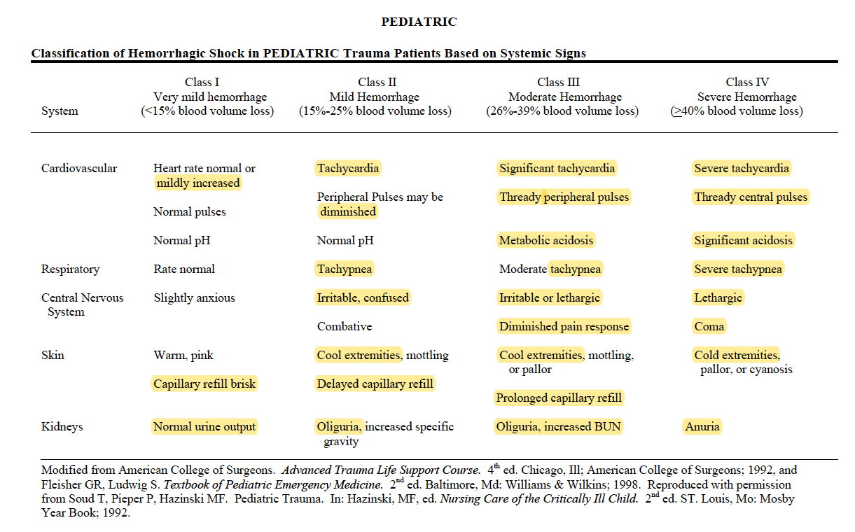
EXAM: ?Dry mucus membrane ?Dry eyes (5% dehydration) ?Sunken fontanelle ?cool peripheries ?oliguria(10%) ?Hypotension ?Tachycardia ?Altered Sensorium (10%) ?prolonged capillary refill time

Naso Gastric tube insertion and 4 hourly aspiration of residue

FLUID RESUSCITATION:

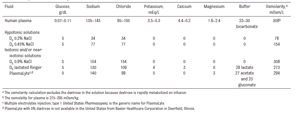
  • For fluid resuscitation : use glucose‑free crystalloids that contain sodium in the range 131–154 mmol/litre, with a bolus of 20 ml/kg over less than 10 minutes. PlasmaLyte, 0.9% Saline ad Ringer Lactate

MAINTENANCE FLUIDS:

  • Calculate routine maintenance IV fluid rates using the Holliday–Segar formula (4 ml/kg/hr for the first 10 kg of weight, 2 ml/kg/hr for the next 10 kg and 1 ml/kg/hr for the weight over 20 kg).
  • This solution would preferably be enriched with glucose 5% (50 ml glucose 50% in 500 ml fluid) in order to provide an adequate caloric supply as recommended (4 to 8 mg glucose/kg/min). In addition, the osmolarity of such a solution makes it possible to be administered on a peripheral venous access.
  • Intraoperative: also will assess for blood loss; the maximum speed of transfusion should be 10ml/kg/hour (if no cardiac failure). Estimated blood volume x ( Desired Hct – Current Hct / Hct of the RBC ) will give the volume of PRBC to be transfused. (N.B.: As a general guide in laparotomies 10 ml/kg/ hour would be needed to compensate for evaporative losses). FFP if needed: 10-15 ml/kg

TARGETS:

  • Normovolemia
  • 1-2 ml/kg/hr of urine OR at least 2 wet nappies
  • Chloride > 95 mmol/L
  • pH <7.5
  • Base excess <6 mmol/L or bicarbonate < 30 mmol/L

CONDUCT OF ANAESTHESIA

  • Ensure that the child is adequately resuscitated
  • Procedure may take 30-60 mins
  • Paediatric anaesthetist
  • Ensure the availability of drugs-equipments for anaesthesia induction, maintenance and resuscitation
  • Attach monitors: ECG, SpO2 and NIBP(AAGBI standards)
  • 4 quadrant aspiration via Ryles tube
  • Preoxygenation for 3 mins
  • Modified rapid sequence induction with cricoid pressure
  • Propofol, Fentanyl and succinyl choline/ rocuronium 1 mg/kg
  • Straight bladed laryngoscope. Insert an uncuffed ETT of size 3.5 mm ID; I will also keep size 3 and 4
  • Ensure proper placement: 5 point auscultation and checking capnography trace
  • Release cricoid pressure and secure the tube
  • Hand ventilate the child before connecting to a ventilator (ventilation guided by ETCO2 values)
  • Paediatric mode; Pressure Controlled Ventilation
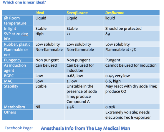
  • O2, Air and sevoflurane maintenance
  • Can consider an epidural for intra and postoperative analgesia (C/I Sepsis, coagulopathy)
  • Local anesthetic can be infiltrated by the surgeon; Ropivacaine or levobupivacaine; dose < 2mg/kg
  • Paracetamol iv (child <10 kg–> 7.5 mg/kg 6 hourly; max 30 mg/kg/day) OR
  • Paracetamol suppository 30 mg/kg loading dose followed by 20 mg/kg 6-8 hourly: max 60 mg/kg/day; continue postoperatively
  • Fentanyl 1microgram/kg iv boluses hourly is less cumulative and more predictable than morphine
  • If well hydrated with good renal perfusion: NSAIDs Ibuprofen 3 mg/kg three times daily or diclofenac per rectal 1 mg/kg/dose suppositories
  • Monitor urine output
  • Antiemetics: ondansetron 0.15 mg/kg
  • Extubation in lateral position
  • Risk of post operative apnoea: SpO2 monitoring with apnoea alarm
  • Regular and PRN analgesics in postoperative period

OTHER INTRAOPERATIVE CONCERNS

  • Strategies to avoid excessive heat loss, optimal analgesia and depth of anaesthesia, optimal fluid therapy
  • Constant monitoring of heart rate, oxygen saturation,EtCO2, blood pressure, body temperature, urine output
  • Check blood sugar to avoid hypoglycemia
  • To prevent hypothermia: Increase the ambient temperature of the OR prior to and during the surgery, body warmer, bubble wraps and plastic drapes to cover the body, cover the head, warm the iv fluids, HME to reduce heat loss from the respiratory system

PAIN EVALUATION IN PAEDIATRICS

  • Evaluated with the support of the parents
  • Using a combination of physiological and behavioural markers in neonates and infants
  • e.g. facial expression, sleeplessness, cry, body movements, posture, increased clinginess, loss of appetite, screaming, reluctance to move, CVS & RS changes
  • Neonate and Infant: Neonatal Pain Agitation and Sedation Score (N-PASS), Neonatal and Infant Pain Scale (NIPS), Face, Legs, Activity Cry and Consolatility scale (FLACC), Objective Pain Score
  • Pre-school and school children can be evaluated by scales like Faces scale ( Happy to crying faces
  • Adolescents: Visual Analogue Scale

POSTOPERATIVE CARE

  • In a high dependency unit or Paediatric Intensive Care unit
  • Will watch for respiratory depression and respiratory compromise, ?shock, pain levels, surgical complications, signs of loss of blood volume
  • Send sample for full blood count and serum electrolytes
  • Continue IV fluids

2.INTUSSUSCEPTION

  • Small bowel telescoping; most common cause of intestinal obstruction in first year of life; other diseases like Meckel’s diverticulum, lymphoma etc can present as intussuception.
  • HISTORY:Present with paroxysmal abdominal pain- child may hold legs to abdomen and red currant jelly like stools. Vomiting and dehydration can cause shock. Abdominal distension can compromise respiration. Infection, infarction,bleeding, perforation, Sepsis-shock etc also can occur.
  • EXAMINATION:Abdomen-guarding. Sausage shaped mass on the right side.
  • INVESTIGATION: Abdominal Ultra sound. X ray abdomen: ?obstruction ?perforation. Air enema can be therapeutic also but contraindicated if having perforation or peritonitis or if in shock.
  • If in shock–> resuscitate and take for laparotomy. A
  • ny repiratory compromise due to distension–> consider the need for mechanical ventilation

3.OESOPHAGEAL ATRESIA (OA) & TRACHEO OESOPHAGEAL FISTULA (TOF)

  • Defective embryonic development of oesophagus and trachea
  • Mostcommon types are 1. isolated OA with distal TOF 2. isolated OA and 3. isolated TOF
  • PAC: ask for SYMPTOMS: Repeated episodes of coughing,chocking and cyanosis worsened by feeding, resitance to pass a nasogastric tube; evaluate for associated congenital anomalies- VACTERL: Vertebral Anal Cardiac Tracheo Esophageal Renal Limb anomalies; problems of prematurity- lung disease, retinal problems, impaired glucose regulation
  • OPTIMISATION: The ligation of a TEF is urgent, but not emergent, except in the setting of respiratory insufficiency severe enough to require ventilatory support. Use IV fluids with glucose to avoid hypoglycemia and to achieve euvolemia. Use specialised suction called Replogle suction to clear the upper part of the oesophagus. Avoiding feeding. Upright positioning of the infant to minimize gastroesophageal reflux. Administration of antibiotic therapy to treat sepsis or aspiration pneumonia. Uncomplicated surgery can be done within 24 hours of birth, to reduce the chance of aspiration of pooled secretions.
  • ANAESTHESIA TECHNIQUE (See above) Specific points for TOF: Under monitoring with pulseoximeter, suction the upper pouch–>Preoxygenation with 100 % O2; avoid vigorous bag and mask ventilation; else the air will pass via TOF into stomach causing distension and splinting of the diaphragm–> An awake technique with titration of small doses of fentanyl (0.2 to 0.5 mcg/kg) allows intubation of the trachea without excessive hemodynamic stimulation or depression. OR an inhalational anesthetic with or without muscle relaxation and with cautious, gentle positive pressure ventilation as needed (Ref: Smith’s anaesthesia for infants & children 8/e) –> place ETT–> allow child to breath spontaneously–>examine airway with a rigid bronchoscope and establish the exact anatomy–> replace and position ETT in a way to occlude the TOF–> once the ETT is placed in the correct position, NMBA can be given. Consider IBP and central venous access based on individual case. Child usually in Right lateral position; take care to maintain the iv access safely.

4.CONGENITAL DIAPHRAGMATIC HERNIA (CDH)

  • Repair is not an emergency procedure. Usually delayed for 24-48 hours for the pulmonary resistance to come down. Baby should be resuscitated first. Delivery should take place as close to term as possible to achieve maximum lung maturity. ABG, Chest x-ray and Echo shold be done. Do an oroogastric suction to clear the part of the bowel in the chest. Intubatin and ventilation should aim to reduce barotrauma with smaller tidal volumes and permissive hypercapnea.

5.EXOMPHALOS & GASTROSCHISIS

  • Due to failure of the migation of the gut into the abdominal cavity during fetal development, there is a herniation of the abdominal contents with a covering sac through a midline defect in the abdominal wall and other congenital anomalies may accompany. Whereas gastroschisis is an isolated anomaly where the gastric contents without any covering sac herniates through a defect in the abdominal wall but not in the midline.
  • There is chance of infection and extensive loss of heat and moisture from the exposed bowel; so it should initially be covered with a non porous material and extreme priority shoud be given for the eveluating the fluid loss and replacement and also for establishing normothermia
  • Primary closure or if respiratory compromise is not allowing it, a staged closure may be necessary

6. INGUINAL HERNIA / REGIONAL TECHNIQUES

  • Mask or IV induction with general anesthesia (with or without endotracheal intubation); spinal anesthesia as a primary anesthetic; caudal anesthesia or analgesia. Muscle relaxation is not necessary but may be a valuable adjunct for decreasing anesthetic requirements and providing optimal surgical conditions.

  • Equipment: Low compression volume anesthesia breathing circuit (circle absorption system vs. Mapleson D ). Monitoring: Standard noninvasive monitoring

  • Maintenance: 1. Inhalation agent plus local infiltration or ilioinguinal-iliohypogastric block on the surgical field OR maintenance caudal analgesia, allow the avoidance of opiates. Neuromuscular blockade with nondepolarizing agent, if chosen. 2. Volume support with judicious amounts of crystalloids.(See 1.) Blood loss is minimal.
  • Emergence and Perioperative Care: 1. Vigilance regarding perioperative abnormalities of control of breathing: periodic breathing/apnea; laryngospasm/bronchospasm; bradycardia; hypoglycemia; continuously monitor with a pulseoximeter. 2. Pain management. (See:1)

Regional anaesthesia:

  • SPINAL ANAESTHESIA: For neonates and infants, a 1 inch 22-gauge spinal needle is inserted at the L4-5 interspace. 0.5 to 0.6 mg/kg of either isobaric or hyperbaric bupivacaine will provide an average of 80 minutes of surgical analgesia for infants and young children who are less than 5 kg in weight. For infants or toddlers 5 to 15 kg, the dosage of hyperbaric bupivacaine or tetracaine is 0.4 mg/kg, and for children weighing more than 15 kg, the dosage of bupivacaine or tetracaine is 0.3 mg/kg . Isobaric ropivacaine has been studied in children for spinal anesthesia at a dosage of 0.5 mg/kg up to 20 mg.
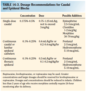
  • CAUDAL BLOCK: The recommended concentration of bupivacaine for a singleshot caudal is 0.125% to 0.25%. An epinephrine dosage of 2.5 mcg/mL, or a concentration of 1:400,000 may be used as an additive for central blocks. Epinephrine will serve as a marker for intravascular injection and decrease systemic absorption of local anesthetic. Additionally, epinephrine may prolong the duration of a regional block. Preservative Free Ketamine, fentanyl, clonidine etc also can also be used as additives. See table: Smith 8/eScreen Shot 2019-08-25 at 11.48.27 am
  • ILIOINGUINAL /ILIOHYPOGASTRIC BLOCK: Can be given after induction, but before the incision; a blunt 22 or 25 gauge needle is inserted 1 cm superior and 1 cm medial to the anterosuperior iliac spine; the needle is initially directed posterolaterally to contact the inner superficial lip of the ileum, then withdrawn while injecting local anesthetic during needle movement. Once skin is reached, the needle is redirected toward the inguinal ligament (ensuring that the needle does not enter the ligament) and local anesthetic is injected after a pop is felt. Can perform under US guidance too. Can use 0.25 mL/kg of 0.5% levobupivacaine or 0.5% ropivacaine at 3 mg/kg. (Ref: Smith’s anaesthesia for infants & children 8/e)
  • Screen Shot 2019-08-29 at 8.18.44 am

VIVA AID: INTRAVENOUS FLUIDS IN PAEDIATRIC POPULATION; SALIENT POINTS FROM VARIOUS GUIDELINES

NICE 2015 GUIDELINES

  • For fluid resuscitation in children and young people: use glucose‑free crystalloids that contain sodium in the range 131–154 mmol/litre, with a bolus of 20 ml/kg over less than 10 minutes. Take into account pre‑existing conditions like cardiac disease or kidney disease, as smaller fluid volumes may be needed.
  • For term neonates who need IV fluid resuscitation: use glucose‑free crystalloids that contain sodium in the range 131–154 mmol/litre, with a bolus of 10–20 ml/kg over less than 10 minutes.
  • Do not use tetrastarch for fluid resuscitation.
  • Calculate routine maintenance IV fluid rates for children and young people using the Holliday–Segar formula (100 ml/kg/day for the first 10 kg of weight, 50 ml/kg/day for the next 10 kg and 20 ml/kg/day for the weight over 20 kg). Be aware that over a 24‑hour period, males rarely need more than 2500 ml and females rarely need more than 2000 ml of fluids.
  • Calculate routine maintenance IV fluid rates for term neonates according to their age, using the following as a guide:
  • From birth to day 1: 50–60 ml/kg/day.
  • Day 2: 70–80 ml/kg/day.
  • Day 3: 80–100 ml/kg/day.
  • Day 4: 100–120 ml/kg/day.
  • Days 5–28: 120–150 ml/kg/day.
  • IV fluids for routine maintenance in children and young people: initially use isotonic crystalloids that contain sodium in the range 131–154 mmol/litre.
  • Measure plasma electrolyte concentrations and blood glucose when starting IV fluids for routine maintenance (except before most elective surgery), and at least every 24 hours thereafter.
  • Base any subsequent IV fluid prescriptions on the plasma electrolyte concentrations and blood glucose measurements.
  • If term neonates need IV fluids for routine maintenance: initially use isotonic crystalloids that contain sodium in the range 131–154 mmol/litre with 5–10% glucose
  • If there is a risk of water retention associated with non‑osmotic antidiuretic hormone (ADH) secretion, consider either:
    • restricting fluids to 50–80% of routine maintenance needs or
    • reducing fluids, calculated on the basis of insensible losses within the range 300–400 ml/m2/24 hours plus urinary output.

Screen Shot 2019-08-16 at 5.28.16 pm

EUROPEAN CONSENSUS 2011 FOR INTRAOPERATIVE FLUID THERAPY

An appropriate solution for intraoperative infusion in children should have an osmolarity and sodium content close to the physiologic range in order to avoid hyponatraemia, an addition of 1–2.5% glucose in order to avoid hypoglycaemia, lipolysis or hyperglycaemia and should also include metabolic anions (i.e. acetate, lactate or malate) as bicarbonate precursors to avoid acid–base balance disturbances (i.e. hyperchloraemic acidosis). The intraoperative infusion of isotonic solutions containing 1–2.5% glucose in children is considered well established use in Europe

BELGIAN PERIOPERATIVE FLUID RECOMMENDATIONS 2012

For children undergoing uncomplicated day-case surgery or minor surgery and those expected to remain nil-by-mouth for at least 24 hours after surgery or undergoing major surgery, a full volume maintenance fluid should be administered during the intraoperative period and immediate post-operative periodas this is associated both with a reduced incidence of postoperative nausea and vomiting particularly in those children receiving opioids, and with a significantly reduced postoperative increase in ADH concentration. The latter presumed to be a result of correction of hypovolemia. 

During the first postoperative day, decreased volumes of the maintenance fluid consisting of an isotonic solution at two-thirds or 70% of the calculated maintenance rate is recommended, provided the child is normovolemic.

This solution would preferably be enriched with glucose 5% (50 ml glucose 50% in 500 ml fluid) in order to provide an adequate caloric supply as recommended (4 to 8 mg glucose/kg/min). In addition, the osmolarity of such a solution makes it possible to be administered on a peripheral venous access.

Recognizing that fluids used to replace ongoing losses should reflect the electrolyte composition of fluid lost, NaCl 0.9% has been considered as appropriate in most cases. Isotonic fluids including colloids are to be used as a bolus in the event of hypovolemia.

Consider iv. fluids as medications.

Administer isotonic fluids (saline 0.9%, Plasmalyte®, Hartmann® or colloids) as a bolus in the event of hypovolemia.

Monitoring plasma electrolytes and glucose concentrations regularly i.e. once daily or more if clinically indicated (documented plasma [Na] < 135 mmol/L)

AMERICAN ACADEMY OF PAEDIATRICS 2018

The American Academy of Pediatrics recommends that patients 28 days to 18 years of age requiring maintenance IVFs should receive isotonic solutions with appropriate potassium chloride and dextrose because they significantly decrease the risk of developing hyponatremia. (For the purposes of this guideline, isotonic solutions have a sodium concentration similar to PlasmaLyte, or 0.9% NaCl)

APA CONSENSUS GUIDELINE ON PERIOPERATIVE FLUID MANAGEMENT IN CHILDREN 2007/ 2010 REVIEW

EXECUTIVE SUMMARY

1. Children can safely be allowed clear fluids 2 hours before surgery without increasing the risk of aspiration.

2. Food should normally be withheld for 6 hours prior to surgery in children aged 6 months or older.

3. In children under 6 months of age it is probably safe to allow a breast milk feed up to 4 hours before surgery

4. Dehydration without signs of hypovolaemia should be corrected slowly.

5. Hypovolaemia should be corrected rapidly to maintain cardiac output and organ perfusion.

6. In the child, a fall in blood pressure is a late sign of hypovolaemia.

7. Maintenance fluid requirements should be calculated using the formula of Holliday and Segar

8. A fluid management plan for any child should address 3 key issues

i. any fluid deficit which is present

ii. maintenance fluid requirements

iii. any losses due to surgery e.g. blood loss, 3rd space losses

9. During surgery all of these requirements should be managed by giving isotonic fluid in all children over 1 month of age

10. The majority of children over 1 month of age will maintain a normal blood sugar if given non-dextrose containing fluid during surgery

11. Children at risk of hypoglycaemia if non-dextrose containing fluid is given are those on parenteral nutrition or a dextrose containing solution prior to theatre, children of low body weight (<3rd centile) or having surgery of more than 3 hours duration and children having extensive regional anaesthesia. These children at risk should be given dextrose containing solutions or have their blood glucose monitored during surgery.

12. Blood loss during surgery should be replaced initially with crystalloid or colloid, and then with blood once the haematocrit has fallen to 25%. Children with cyanotic congenital heart disease and neonates may need a higher haematocrit to maintain oxygenation.

13. Fluid therapy should be monitored by daily electrolyte estimation, use of a fluid input/output chart and daily weighing if feasible.

14. Acute dilutional hyponatraemia is a medical emergency and should be managed in PICU.