VIVA SCENE IMAGING: CHEST X-RAY (CXR)

  • Helpful MNEMONICS: ‘PRIP ABCDEFGHI’. Also in inspiratory films the level of the diaphragm is at the level of ribs 5/6 anteriorly and 8/10 posteriorly. (AR6PR10). 
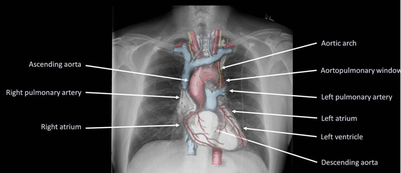
  • Right heart border is formed predominantly by the right atrium along with the lower part of SVC, whilst the left border is formed by the aortic arch, pulmonary artery, left atrium, and ventricle. The right and left ventricle forms the inferior border. The right diaphragm is normally higher than the left due to the liver.
  • Areas where pathology is commonly missed: Apices (including behind the 1st rib and clavicle)—small pneumothoraces and masses. Hila—masses and lymph nodes; left hilum is 1–2 cm higher than right. Behind the heart—left lower lobar collapse and hiatus hernia. Below the diaphragm—free gas. Soft tissues—breast shadow or absence (look for lung and bone metastasis)
  • This is CXR of Mr Roy (Patient identity) done on 14/08/2019 (date of study)
  • Its an AP film (Projection)
  • Non rotated (Rotation)
  • Expiratory film (Inspiratory/Expiratory)
  • Adequately penetrated (Penetrated)
  • Trachea is central, no foreign bodies or other abnormalities (A– Airway)
  • On both sides, the bones and soft tissues appear normal; ribs, sternum, scapula, clavicle, spine, and humerus has no deposits or fractures (B-Bones and soft tissues)
  • The cardiac silhoutte is normal (C-Cardiac silhoutte)
  • There is no free air under the diaphragm. Bilateral costo- and cardio-phrenic angles are clear (D– Diaphragm and angles)
  • No effusions. Pleura is not visible (E-Effusion)
  • Both sides, the lung fields appear normal (F-Fields = lung fields)
  • Gastric bubble visible at left upper abdomen (G=Gastric bubble)
  • The hila and the mediastenum appears to be normal and not displaced. The CT ratio is less than 50% (H-Hila and mediastenum)
  • Endotracheal tube tip is seen 5 cms above the carina or T4/T5 interspace. NG tube passed down midline, past level of diaphragm, and deviates to left with tip seen in stomach. ECG leads and electrodes are noted (I=Insertions, Interventions: tubes and lines, chest drains, pacemakers, and metallic valves and artefacts)
Screen Shot 2019-08-25 at 5.10.08 pm
  • If an intrathoracic opacity is in anatomical contact with the heart border, then the opacity will obscure that border. If an intrathoracic opacity is in the posterior pleural cavity so not in direct anatomical contact with the heart border, this causes an overlap but not an obliteration of that border (Silhoutte sign)
  • When the alveoli no longer contain air and opacify, the air-filled bronchi passing through the alveoli may be visible as branching linear lucencies. (Air bronchogram)
  • Linear opacities measuring 1–6 cm extending from periphery to the hila caused by distension of anastomotic channels between peripheral and central lymphatics.(Kerley A lines)
  • Short horizontal lines, due to oedema of the interlobular septae, situated perpendicularly to the pleural surface at the lung base (Kerley B lines)
  • Reticular opacities at the lung base (Kerley C lines)
  • The mediastinal masses can be identified on the PA views but lateral films and/or CT scans are required to confirm further diagnosis
  • Anterior mediastenum: Anterior to trachea, Middle mediastenum: Anterior to heart, Posterior mediastenum: Posterior to heart
  • ANTERIOR MEDIASTENAL MASS: (4 T’s) Thymic tumour -Teratoma- Thyroid –  Terrible lymph nodes (LN) Ascending aortic aneurysm, MIDDLE MEDIASTENAL MASS (BAL) Bronchogenic cyst – Arch aneurysm- LNs, POSTERIOR MEDIASTENAL MASS: as expected it can be from Spine/Neurogenic; also hiatus hernia
  • Hilar mass: Most commonly TB/Sarcoidosis; also lymphoma
  • Collapse: Triangular opacity, Crowding of ribs, Mediastenal displacement, hyperinflation of other lobes
  • Consolidation: confluent ill defined opacity, bat wing distribution, no volume loss, air bronchogram
  • Effusion: Erect: obliteration of the costo- and cardio-phrenic angles and opacity with meniscus. Supine: graded haze giving a ground glass opacity
  • Emphysema: Decreased lung markings, flat diaphragm and hyperexpanded lung, Presence of bullae and peribronchial thickening, signs of cor pulmonale, right ventricular enlargement, pulmonary hypertension—enlargement of the central pulmonary arteries with oligaemic peripheral lung fields

VIVA SCENE : NEONATE /OLDER CHILD Vs ADULT

CVS:

  • Average HR is 120-180 bpm and SBP 50-90 mm of Hg at birth
  • Oxygen consumption is twice that of an adult 7 ml/kg/min. But because of a fixed stroke volume, increasing the HR is the only way to increase CO. Also neonatal myocardium consists of more non-contractile connective tissue.
  • Parasympathetic system is predominant; bradycardia can happen in response to hypoxia or tracheal suction. Asystole is the most common form of cardiac arrest and ventricular fibrillation is uncommon
  • Circulating volume approx 90 ml/kg @ birth (300-400 ml in an average neonate). Bleeding contributes proportionately a greater loss of total volume in a neonate, compared to an adult. Right ventricular mass equal to left ventricular mass until 6 months of age, resulting in right axis deviation on the ECG.
  • Transitional circulation: Before birth the SVR is low due to the low resistance placental circulation whereas the pulmonary resistance is high. At birth the SVR rapidly increases after clamping of the umbilical cord and PVR decreases following functional closure of foramen ovale (due to increased venous return to left atrium from pulmonary arteries) and ductus arteriosus (in response to increased PaO2 in blood). But this can be reversed back to the foetal state due to stimuli including hypoxia, heypecarbia and acidosis, leading to a perpetuating cycle of worsening hypoxia. This is known as transitional circulation or prsistent foetal circulation.

AIRWAY/ RESPIRATORY SYSTEM

  • Relatively larger head, short neck, large tongue and narrow nasal passages.
  • High anterior larynx (level C2/3 compared to C5/6 in the adult).
  • Large U-shaped floppy epiglottis.
  • Narrowest point of the larynx is at the level of the cricoid cartilage (in adults it is at the laryngeal inlet).
  • Trauma to the small airway can easily lead to oedema and airway obstruction. 1 mm oedema can narrow an infant’s airway by 60% (resistance ∝1/radius).
  • Equal angles of mainstem bronchi (in adults the right main bronchus is more vertical).
  • A compliant chestwall: FRC is less as the elastic recoil pulls the compliant chest wall inwards. Closing volume is larger than FRC until 6–8 years of age resulting in airway closure at end-expiration; this can be reduced by CPAP
  • Fatiguable respiratory and accessory muscles: The diaphragm has less % of fatigue resistant type I respiratory fibres. Raised abdominal pressure can splint the diaphragm precipitating respiratory failure. Diaphragmatic breathing > intercostal breathing. Diaphragmatic movement restricted by relatively large liver.
  • Horizontally aligned ribs prevent the bucket handle movement of the ribs during inspiration
  • Incompletely developed alveoli.Born with only 10% of the total number of alveoli as adults. Alveoli develop over first 8 years. Higher alveolar ventilation 100–150 mL/kg/min compared with 60 mL/kg/min in adult.
  • Inconsistant ventilatory response to hypercapnoea. Higher risk of apnoea
  • Sinusoidal respiratory pattern, no end-expiratory pause (inspiratory/expiratory ratio 1:1). Resting repiratory rate is high and increases further if respiratory compensation is required. Limited capability to increase the tidal volume
  • Obligate nasal breathers; nasal obstruction (e.g. choanal atresia, respiratory infection) can precipitate respiratory failure
  • MAC infant > neonate > adult

CNS

  • CNS development is incomplete at birth
  • Myelination starts before birth and continues throughout the first year
  • Spinal cord ends at L3 (L1 by age 2 years).
  • Unfused fontanelle and sutures makes it more compliant and can expand to some extent in response to raised ICP. ICP is less than the adult: 2-4 mm of Hg
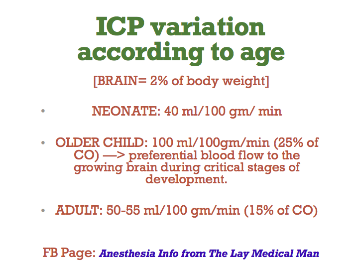
  • Cerebral Blood Flow is less in the neonate than the adult and autoregulation operates at less SBPs
  • In premature infants autoregulation is absent and perfusion is pressure dependent
  • Blood brain barrier is more permeable and hence the neonate is more sensitive to sedatives
  • Neonate responds to pain with tachycardia, hypertension, grimaces etc

Screen Shot 2019-08-24 at 12.17.29 pm

TEMPERATURE REGULATION

  • Higher thermoneutral temperature (temperature below which an individual is unable to maintain core body temperature) 32 degree C for a term infant compared with 28 for an adult.

  • Neonate loss heat very readily due to a high body surface area to weight ratio and absence of shivering in infants ❤ months
  • Produces heat by metabolism of brown fat (5-6% of body wt, stored around kidneys, scapula and mediastenum) by non shivering thermogenesis. Beta adrenergic mediated and contributes to significant amount of the O2 demand placed on the cardiorespiratory system. Ablated by beta blockade

RENAL SYSTEM

  • Immature at birth
  • Higher total body water (80%) at birth
  • Renal blood flow is 6% of cardiac output at birth rising to 18% of cardiac output at 1 month (compared with 20% in adult).
  • Reduced GFR, tubular function until 6–8 months of age
  • Reduced H+ ion excretion
  • Cannot tolerate both excessive water/ sodium load and dehydration (decresed ability to conserve water)
  • Fluids must be carefully balanced based on weight, insensible and observed fluid losses and maintenance requirements

HEPATIC SYSTEM

Low hepatic glycogen stores means hypoglycaemia occurs readily with prolonged fasting

DRUG ADMINISTRATION

  • TBW is increased: so the volume of distribution of water soluble drugs will increase and hence need an increase in the dose. e.g. Succinyl choline (1 mg/kg in adult but 2 mg/kg in the neonate)
  • Succinyl choline by acting on the SA node can produce arrhythmias and even asystole in the neonate
  • Though the neonate is more sensitive to nondepolarizing muscle relaxants, the higher ECF volume increases the required dose; so the final required dose remains unchanged
  • Immature hepatic and renal systems make the metabolism and excretion of the drugs slow. e.g. half life of morphine is increased due to the reduced ability to produce glucoronide conjugates
  • Circulating levels of albumin and alpha acid glycoprotein is less, increasing the free fraction and a pronounced effect
  • MAC is age related. MAC values for an infant are: • sevoflurane = 3.3% • isoflurane = 1.9% • desflurane = 9.4%.

VIVA SCENE: ALL IMPORTANT PAEDIATRIC CASES IN ONE PLACE

1. PYLORIC STENOSIS (PROTOTYPE CASE; ANY OTHER CASE, MAKE THIS A TEMPLATE AND ADD SPECIFIC POINTS RELEVANT FOR THAT CONDITION)

Most common cause of intestinal obstruction in infancy

Due to hypertrophy of circular pyloric muscle

Presentation: 4-6 weeks of age

Persistent projectile vomiting

There is obstruction at the level of pylorus: so bicarbonate rich fluid from intestine cannot mix with gastric secretions

So the vomiting causes metabolic alkalosis and hypokalemia due to loss of acidic gastric juice alone
The large bicarbonate load is in excess of kidney’s absorptive capacity and the urine becomes alkaline initially
Later when the fluid and electrolyte loss result in dehydration, the renin angiotensin system is activated and result in aldosterone secretion which tries to preserve sodium at the expense of K and Cl ions

This result in production of paradoxical acidic urine and worsening of metabolic alkalosis and hypokalemia

An attempt to compensate it through hypoventilation is initiated; but it will be insufficient and also this will trigger the hypoxic drive!

Hypoglycemia, hemoconcentration, mild uremia and unconjugated hyperbilirubinemia may be seen.
ANAESTHETIC CONCERNS

Pyloric stenosis is not an emergency

The DYSELECTROLYTEMIA and ACID BASE IMBALANCE should be corrected before taking up for surgery.

Increased chance for regurgitation, altered physiology and anatomy, altered drug dosages, difficult venous access, anxious parents are other concerns

There should be an experienced paediatric anaesthesiologist for the conduct of anaesthesia
P.A.C. AND EVALUATION

LOCATION: Needs resuscitation in a paediatric ICU or ward

INVESTIGATIONS: CBC, Blood sugar, RFT, LFT, group and hold, serial ABGs to know the effectiveness of resuscitation and decide on the dose of K administration. 3 mmol/kg/24 hour potassium should be added to maintenance fluids

HISTORY: From the parent; vomiting frequency, amount of feeds taken, diarrhoea, frequency of wetting of the nappy, fever, altered sensorium

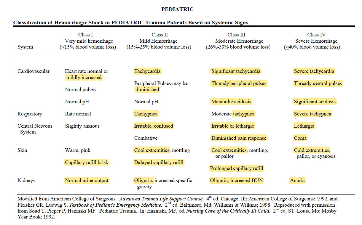
EXAM: ?Dry mucus membrane ?Dry eyes (5% dehydration) ?Sunken fontanelle ?cool peripheries ?oliguria(10%) ?Hypotension ?Tachycardia ?Altered Sensorium (10%) ?prolonged capillary refill time

Naso Gastric tube insertion and 4 hourly aspiration of residue

FLUID RESUSCITATION:

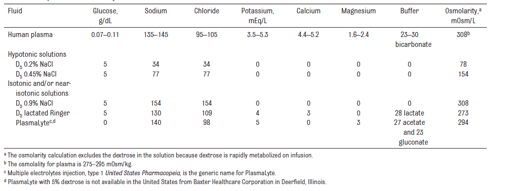
  • For fluid resuscitation : use glucose‑free crystalloids that contain sodium in the range 131–154 mmol/litre, with a bolus of 20 ml/kg over less than 10 minutes. PlasmaLyte, 0.9% Saline ad Ringer Lactate

MAINTENANCE FLUIDS:

  • Calculate routine maintenance IV fluid rates using the Holliday–Segar formula (4 ml/kg/hr for the first 10 kg of weight, 2 ml/kg/hr for the next 10 kg and 1 ml/kg/hr for the weight over 20 kg).
  • This solution would preferably be enriched with glucose 5% (50 ml glucose 50% in 500 ml fluid) in order to provide an adequate caloric supply as recommended (4 to 8 mg glucose/kg/min). In addition, the osmolarity of such a solution makes it possible to be administered on a peripheral venous access.
  • Intraoperative: also will assess for blood loss; the maximum speed of transfusion should be 10ml/kg/hour (if no cardiac failure). Estimated blood volume x ( Desired Hct – Current Hct / Hct of the RBC ) will give the volume of PRBC to be transfused. (N.B.: As a general guide in laparotomies 10 ml/kg/ hour would be needed to compensate for evaporative losses). FFP if needed: 10-15 ml/kg

TARGETS:

  • Normovolemia
  • 1-2 ml/kg/hr of urine OR at least 2 wet nappies
  • Chloride > 95 mmol/L
  • pH <7.5
  • Base excess <6 mmol/L or bicarbonate < 30 mmol/L

CONDUCT OF ANAESTHESIA

  • Ensure that the child is adequately resuscitated
  • Procedure may take 30-60 mins
  • Paediatric anaesthetist
  • Ensure the availability of drugs-equipments for anaesthesia induction, maintenance and resuscitation
  • Attach monitors: ECG, SpO2 and NIBP(AAGBI standards)
  • 4 quadrant aspiration via Ryles tube
  • Preoxygenation for 3 mins
  • Modified rapid sequence induction with cricoid pressure
  • Propofol, Fentanyl and succinyl choline/ rocuronium 1 mg/kg
  • Straight bladed laryngoscope. Insert an uncuffed ETT of size 3.5 mm ID; I will also keep size 3 and 4
  • Ensure proper placement: 5 point auscultation and checking capnography trace
  • Release cricoid pressure and secure the tube
  • Hand ventilate the child before connecting to a ventilator (ventilation guided by ETCO2 values)
  • Paediatric mode; Pressure Controlled Ventilation
  • O2, Air and sevoflurane maintenance
  • Can consider an epidural for intra and postoperative analgesia (C/I Sepsis, coagulopathy)
  • Local anesthetic can be infiltrated by the surgeon; Ropivacaine or levobupivacaine; dose < 2mg/kg
  • Paracetamol iv (child <10 kg–> 7.5 mg/kg 6 hourly; max 30 mg/kg/day) OR
  • Paracetamol suppository 30 mg/kg loading dose followed by 20 mg/kg 6-8 hourly: max 60 mg/kg/day; continue postoperatively
  • Fentanyl 1microgram/kg iv boluses hourly is less cumulative and more predictable than morphine
  • If well hydrated with good renal perfusion: NSAIDs Ibuprofen 3 mg/kg three times daily or diclofenac per rectal 1 mg/kg/dose suppositories
  • Monitor urine output
  • Antiemetics: ondansetron 0.15 mg/kg
  • Extubation in lateral position
  • Risk of post operative apnoea: SpO2 monitoring with apnoea alarm
  • Regular and PRN analgesics in postoperative period

OTHER INTRAOPERATIVE CONCERNS

  • Strategies to avoid excessive heat loss, optimal analgesia and depth of anaesthesia, optimal fluid therapy
  • Constant monitoring of heart rate, oxygen saturation,EtCO2, blood pressure, body temperature, urine output
  • Check blood sugar to avoid hypoglycemia
  • To prevent hypothermia: Increase the ambient temperature of the OR prior to and during the surgery, body warmer, bubble wraps and plastic drapes to cover the body, cover the head, warm the iv fluids, HME to reduce heat loss from the respiratory system

PAIN EVALUATION IN PAEDIATRICS

  • Evaluated with the support of the parents
  • Using a combination of physiological and behavioural markers in neonates and infants
  • e.g. facial expression, sleeplessness, cry, body movements, posture, increased clinginess, loss of appetite, screaming, reluctance to move, CVS & RS changes
  • Neonate and Infant: Neonatal Pain Agitation and Sedation Score (N-PASS), Neonatal and Infant Pain Scale (NIPS), Face, Legs, Activity Cry and Consolatility scale (FLACC), Objective Pain Score
  • Pre-school and school children can be evaluated by scales like Faces scale ( Happy to crying faces
  • Adolescents: Visual Analogue Scale

POSTOPERATIVE CARE

  • In a high dependency unit or Paediatric Intensive Care unit
  • Will watch for respiratory depression and respiratory compromise, ?shock, pain levels, surgical complications, signs of loss of blood volume
  • Send sample for full blood count and serum electrolytes
  • Continue IV fluids

2.INTUSSUSCEPTION

  • Small bowel telescoping; most common cause of intestinal obstruction in first year of life; other diseases like Meckel’s diverticulum, lymphoma etc can present as intussuception.
  • HISTORY:Present with paroxysmal abdominal pain- child may hold legs to abdomen and red currant jelly like stools. Vomiting and dehydration can cause shock. Abdominal distension can compromise respiration. Infection, infarction,bleeding, perforation, Sepsis-shock etc also can occur.
  • EXAMINATION:Abdomen-guarding. Sausage shaped mass on the right side.
  • INVESTIGATION: Abdominal Ultra sound. X ray abdomen: ?obstruction ?perforation. Air enema can be therapeutic also but contraindicated if having perforation or peritonitis or if in shock.
  • If in shock–> resuscitate and take for laparotomy. A
  • ny repiratory compromise due to distension–> consider the need for mechanical ventilation

3.OESOPHAGEAL ATRESIA (OA) & TRACHEO OESOPHAGEAL FISTULA (TOF)

  • Defective embryonic development of oesophagus and trachea
  • Mostcommon types are 1. isolated OA with distal TOF 2. isolated OA and 3. isolated TOF
  • PAC: ask for SYMPTOMS: Repeated episodes of coughing,chocking and cyanosis worsened by feeding, resitance to pass a nasogastric tube; evaluate for associated congenital anomalies- VACTERL: Vertebral Anal Cardiac Tracheo Esophageal Renal Limb anomalies; problems of prematurity- lung disease, retinal problems, impaired glucose regulation
  • OPTIMISATION: The ligation of a TEF is urgent, but not emergent, except in the setting of respiratory insufficiency severe enough to require ventilatory support. Use IV fluids with glucose to avoid hypoglycemia and to achieve euvolemia. Use specialised suction called Replogle suction to clear the upper part of the oesophagus. Avoiding feeding. Upright positioning of the infant to minimize gastroesophageal reflux. Administration of antibiotic therapy to treat sepsis or aspiration pneumonia. Uncomplicated surgery can be done within 24 hours of birth, to reduce the chance of aspiration of pooled secretions.
  • ANAESTHESIA TECHNIQUE (See above) Specific points for TOF: Under monitoring with pulseoximeter, suction the upper pouch–>Preoxygenation with 100 % O2; avoid vigorous bag and mask ventilation; else the air will pass via TOF into stomach causing distension and splinting of the diaphragm–> An awake technique with titration of small doses of fentanyl (0.2 to 0.5 mcg/kg) allows intubation of the trachea without excessive hemodynamic stimulation or depression. OR an inhalational anesthetic with or without muscle relaxation and with cautious, gentle positive pressure ventilation as needed (Ref: Smith’s anaesthesia for infants & children 8/e) –> place ETT–> allow child to breath spontaneously–>examine airway with a rigid bronchoscope and establish the exact anatomy–> replace and position ETT in a way to occlude the TOF–> once the ETT is placed in the correct position, NMBA can be given. Consider IBP and central venous access based on individual case. Child usually in Right lateral position; take care to maintain the iv access safely.

4.CONGENITAL DIAPHRAGMATIC HERNIA (CDH)

  • Repair is not an emergency procedure. Usually delayed for 24-48 hours for the pulmonary resistance to come down. Baby should be resuscitated first. Delivery should take place as close to term as possible to achieve maximum lung maturity. ABG, Chest x-ray and Echo shold be done. Do an oroogastric suction to clear the part of the bowel in the chest. Intubatin and ventilation should aim to reduce barotrauma with smaller tidal volumes and permissive hypercapnea.

5.EXOMPHALOS & GASTROSCHISIS

  • Due to failure of the migation of the gut into the abdominal cavity during fetal development, there is a herniation of the abdominal contents with a covering sac through a midline defect in the abdominal wall and other congenital anomalies may accompany. Whereas gastroschisis is an isolated anomaly where the gastric contents without any covering sac herniates through a defect in the abdominal wall but not in the midline.
  • There is chance of infection and extensive loss of heat and moisture from the exposed bowel; so it should initially be covered with a non porous material and extreme priority shoud be given for the eveluating the fluid loss and replacement and also for establishing normothermia
  • Primary closure or if respiratory compromise is not allowing it, a staged closure may be necessary

6. INGUINAL HERNIA / REGIONAL TECHNIQUES

  • Mask or IV induction with general anesthesia (with or without endotracheal intubation); spinal anesthesia as a primary anesthetic; caudal anesthesia or analgesia. Muscle relaxation is not necessary but may be a valuable adjunct for decreasing anesthetic requirements and providing optimal surgical conditions.

  • Equipment: Low compression volume anesthesia breathing circuit (circle absorption system vs. Mapleson D ). Monitoring: Standard noninvasive monitoring

  • Maintenance: 1. Inhalation agent plus local infiltration or ilioinguinal-iliohypogastric block on the surgical field OR maintenance caudal analgesia, allow the avoidance of opiates. Neuromuscular blockade with nondepolarizing agent, if chosen. 2. Volume support with judicious amounts of crystalloids.(See 1.) Blood loss is minimal.
  • Emergence and Perioperative Care: 1. Vigilance regarding perioperative abnormalities of control of breathing: periodic breathing/apnea; laryngospasm/bronchospasm; bradycardia; hypoglycemia; continuously monitor with a pulseoximeter. 2. Pain management. (See:1)

Regional anaesthesia:

  • SPINAL ANAESTHESIA: For neonates and infants, a 1 inch 22-gauge spinal needle is inserted at the L4-5 interspace. 0.5 to 0.6 mg/kg of either isobaric or hyperbaric bupivacaine will provide an average of 80 minutes of surgical analgesia for infants and young children who are less than 5 kg in weight. For infants or toddlers 5 to 15 kg, the dosage of hyperbaric bupivacaine or tetracaine is 0.4 mg/kg, and for children weighing more than 15 kg, the dosage of bupivacaine or tetracaine is 0.3 mg/kg . Isobaric ropivacaine has been studied in children for spinal anesthesia at a dosage of 0.5 mg/kg up to 20 mg.
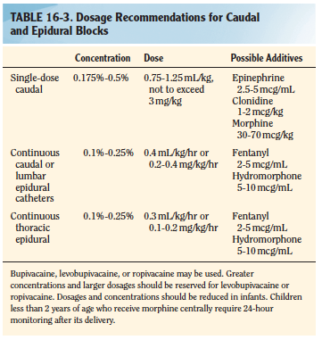
  • CAUDAL BLOCK: The recommended concentration of bupivacaine for a singleshot caudal is 0.125% to 0.25%. An epinephrine dosage of 2.5 mcg/mL, or a concentration of 1:400,000 may be used as an additive for central blocks. Epinephrine will serve as a marker for intravascular injection and decrease systemic absorption of local anesthetic. Additionally, epinephrine may prolong the duration of a regional block. Preservative Free Ketamine, fentanyl, clonidine etc also can also be used as additives. See table: Smith 8/eScreen Shot 2019-08-25 at 11.48.27 am
  • ILIOINGUINAL /ILIOHYPOGASTRIC BLOCK: Can be given after induction, but before the incision; a blunt 22 or 25 gauge needle is inserted 1 cm superior and 1 cm medial to the anterosuperior iliac spine; the needle is initially directed posterolaterally to contact the inner superficial lip of the ileum, then withdrawn while injecting local anesthetic during needle movement. Once skin is reached, the needle is redirected toward the inguinal ligament (ensuring that the needle does not enter the ligament) and local anesthetic is injected after a pop is felt. Can perform under US guidance too. Can use 0.25 mL/kg of 0.5% levobupivacaine or 0.5% ropivacaine at 3 mg/kg. (Ref: Smith’s anaesthesia for infants & children 8/e)
  • Screen Shot 2019-08-29 at 8.18.44 am

VIVA AID: INTRAVENOUS FLUIDS IN PAEDIATRIC POPULATION; SALIENT POINTS FROM VARIOUS GUIDELINES

NICE 2015 GUIDELINES

  • For fluid resuscitation in children and young people: use glucose‑free crystalloids that contain sodium in the range 131–154 mmol/litre, with a bolus of 20 ml/kg over less than 10 minutes. Take into account pre‑existing conditions like cardiac disease or kidney disease, as smaller fluid volumes may be needed.
  • For term neonates who need IV fluid resuscitation: use glucose‑free crystalloids that contain sodium in the range 131–154 mmol/litre, with a bolus of 10–20 ml/kg over less than 10 minutes.
  • Do not use tetrastarch for fluid resuscitation.
  • Calculate routine maintenance IV fluid rates for children and young people using the Holliday–Segar formula (100 ml/kg/day for the first 10 kg of weight, 50 ml/kg/day for the next 10 kg and 20 ml/kg/day for the weight over 20 kg). Be aware that over a 24‑hour period, males rarely need more than 2500 ml and females rarely need more than 2000 ml of fluids.
  • Calculate routine maintenance IV fluid rates for term neonates according to their age, using the following as a guide:
  • From birth to day 1: 50–60 ml/kg/day.
  • Day 2: 70–80 ml/kg/day.
  • Day 3: 80–100 ml/kg/day.
  • Day 4: 100–120 ml/kg/day.
  • Days 5–28: 120–150 ml/kg/day.
  • IV fluids for routine maintenance in children and young people: initially use isotonic crystalloids that contain sodium in the range 131–154 mmol/litre.
  • Measure plasma electrolyte concentrations and blood glucose when starting IV fluids for routine maintenance (except before most elective surgery), and at least every 24 hours thereafter.
  • Base any subsequent IV fluid prescriptions on the plasma electrolyte concentrations and blood glucose measurements.
  • If term neonates need IV fluids for routine maintenance: initially use isotonic crystalloids that contain sodium in the range 131–154 mmol/litre with 5–10% glucose
  • If there is a risk of water retention associated with non‑osmotic antidiuretic hormone (ADH) secretion, consider either:
    • restricting fluids to 50–80% of routine maintenance needs or
    • reducing fluids, calculated on the basis of insensible losses within the range 300–400 ml/m2/24 hours plus urinary output.

Screen Shot 2019-08-16 at 5.28.16 pm

EUROPEAN CONSENSUS 2011 FOR INTRAOPERATIVE FLUID THERAPY

An appropriate solution for intraoperative infusion in children should have an osmolarity and sodium content close to the physiologic range in order to avoid hyponatraemia, an addition of 1–2.5% glucose in order to avoid hypoglycaemia, lipolysis or hyperglycaemia and should also include metabolic anions (i.e. acetate, lactate or malate) as bicarbonate precursors to avoid acid–base balance disturbances (i.e. hyperchloraemic acidosis). The intraoperative infusion of isotonic solutions containing 1–2.5% glucose in children is considered well established use in Europe

BELGIAN PERIOPERATIVE FLUID RECOMMENDATIONS 2012

For children undergoing uncomplicated day-case surgery or minor surgery and those expected to remain nil-by-mouth for at least 24 hours after surgery or undergoing major surgery, a full volume maintenance fluid should be administered during the intraoperative period and immediate post-operative periodas this is associated both with a reduced incidence of postoperative nausea and vomiting particularly in those children receiving opioids, and with a significantly reduced postoperative increase in ADH concentration. The latter presumed to be a result of correction of hypovolemia. 

During the first postoperative day, decreased volumes of the maintenance fluid consisting of an isotonic solution at two-thirds or 70% of the calculated maintenance rate is recommended, provided the child is normovolemic.

This solution would preferably be enriched with glucose 5% (50 ml glucose 50% in 500 ml fluid) in order to provide an adequate caloric supply as recommended (4 to 8 mg glucose/kg/min). In addition, the osmolarity of such a solution makes it possible to be administered on a peripheral venous access.

Recognizing that fluids used to replace ongoing losses should reflect the electrolyte composition of fluid lost, NaCl 0.9% has been considered as appropriate in most cases. Isotonic fluids including colloids are to be used as a bolus in the event of hypovolemia.

Consider iv. fluids as medications.

Administer isotonic fluids (saline 0.9%, Plasmalyte®, Hartmann® or colloids) as a bolus in the event of hypovolemia.

Monitoring plasma electrolytes and glucose concentrations regularly i.e. once daily or more if clinically indicated (documented plasma [Na] < 135 mmol/L)

AMERICAN ACADEMY OF PAEDIATRICS 2018

The American Academy of Pediatrics recommends that patients 28 days to 18 years of age requiring maintenance IVFs should receive isotonic solutions with appropriate potassium chloride and dextrose because they significantly decrease the risk of developing hyponatremia. (For the purposes of this guideline, isotonic solutions have a sodium concentration similar to PlasmaLyte, or 0.9% NaCl)

APA CONSENSUS GUIDELINE ON PERIOPERATIVE FLUID MANAGEMENT IN CHILDREN 2007/ 2010 REVIEW

EXECUTIVE SUMMARY

1. Children can safely be allowed clear fluids 2 hours before surgery without increasing the risk of aspiration.

2. Food should normally be withheld for 6 hours prior to surgery in children aged 6 months or older.

3. In children under 6 months of age it is probably safe to allow a breast milk feed up to 4 hours before surgery

4. Dehydration without signs of hypovolaemia should be corrected slowly.

5. Hypovolaemia should be corrected rapidly to maintain cardiac output and organ perfusion.

6. In the child, a fall in blood pressure is a late sign of hypovolaemia.

7. Maintenance fluid requirements should be calculated using the formula of Holliday and Segar

8. A fluid management plan for any child should address 3 key issues

i. any fluid deficit which is present

ii. maintenance fluid requirements

iii. any losses due to surgery e.g. blood loss, 3rd space losses

9. During surgery all of these requirements should be managed by giving isotonic fluid in all children over 1 month of age

10. The majority of children over 1 month of age will maintain a normal blood sugar if given non-dextrose containing fluid during surgery

11. Children at risk of hypoglycaemia if non-dextrose containing fluid is given are those on parenteral nutrition or a dextrose containing solution prior to theatre, children of low body weight (<3rd centile) or having surgery of more than 3 hours duration and children having extensive regional anaesthesia. These children at risk should be given dextrose containing solutions or have their blood glucose monitored during surgery.

12. Blood loss during surgery should be replaced initially with crystalloid or colloid, and then with blood once the haematocrit has fallen to 25%. Children with cyanotic congenital heart disease and neonates may need a higher haematocrit to maintain oxygenation.

13. Fluid therapy should be monitored by daily electrolyte estimation, use of a fluid input/output chart and daily weighing if feasible.

14. Acute dilutional hyponatraemia is a medical emergency and should be managed in PICU.

VIVA SCENE: Neuroanaesthesia- Management of Traumatic Brain Injury and other questions

TBI MANAGEMENT (Based on BTF 4e Guidelines)

  • Principal of management of head injury is to prevent secondary brain injury due to hypoxia, hyper/hypocarbia, hypovolaemia, hypotension, and increased ICP.
  • Primary survey and management of other life-threatening injury (tension pneumothorax, cardiac tamponade, airway obstruction, etc.) assessment with GCS
  • Continuous high-flow O2 for all potential TBI cases
  • Airway repositioning maneuvers if needed
  • Evaluate the GCS. GCS<8, increased risk of aspiration, concern for hypoxemia and hypercarbia, need for hyperventilation: all such concerns when present are addressed with intubation and mechanical ventilation.
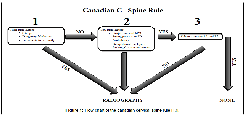
  • C-Spine imaging based on NEXUS or Canadian C Spine rule criteria (see below)
  • Obtain large bore IV’s and/or central venous access for patients requiring vasoactive drugs. For surgical procedures:
  • ASA monitors along with direct intra-arterial pressure monitoring (zeroed at the level of the head to facilitate assessment of cerebral perfusion pressure [CPP]) and bladder catheterization are established. Avoid hypotension due to induction drugs and surges in BP during intubation and pinning using slow drug administration and vasoactive drugs.
  • All attempts at intubation should include in-line neck stabilization. Anesthetic drugs that allow for rapid control of the airway while avoiding an increase in intracranial pressure (ICP) and providing hemodynamic stability are preferred. For rapid sequence intubation, succinylcholine or rocuronium may be used.
  • Choice of drugs must be tailored to each individual patient. IV anesthetics are cerebral vasoconstrictors whereas volatile agents increase cerebral blood flow (CBF) above 1 MAC
  • Lidocaine in doses of 1.5 mg/kg may inhibit the adverse effects of laryngoscopy decreasing ICP, CMR, and CBF with minimal hemodynamic effects

  • Careful positioning to avoid impedance to venous drainage due to extreme neck rotation or tilt. Avoid ties around the neck for endotracheal tube fixation
  • Avoid increases in intrathoracic pressure (obstructed endotracheal tubes, bronchospasm).
  • Maintain SBP at ≥ 110 mmHg  for patients 15 to 49 or over 70 years and at ≥ 100 mmHg for patients 50 to 69years old. The recommended target CPP value is between 60 and 70 mmHg
  • Isotonic crystalloid is the preferred fluid..A single SBP measurement < 90 mmHg will initiate intravenous (IV) fluid resuscitation with an initial bolus of 1 L of NS/RL in adults and adolescents and 20 ml/kg in older children followed by maintenance rates to keep SBP ≥ 90 mmHg
  • Also hypovolemia resulting from extracranial hemorrhage should be ruled out
  • Maintain ETCO2 between 35 and 45 mmHg (4.5-6 kPa). Avoid hyperventilation, especially in the first 12 hoursIf you are going to hyperventilate the patient, use jugular venous saturation monitoring to ensure the brain is getting enough oxygen
  • ABGs to titrate the ventilation and manage the fluids and electrolytes administration and to decide on postoperative ventilation if needed
  • Avoid hypo and hyperglycemia
  • Anaesthetic technique should allow a smooth and rapid recovery and prompt neurological assessment. Again avoid bucking over the endotracheal tube. Labetalol can be used to control sympathetic surges during extubation
  • Continuous monitoring to avoid hypovolemia, hypotension, hypercarbia, hypoxia, hypoglycemia and dyselectrolytemias post extubation
  • Adequate perioperative analgesia is important to prevent further raise in ICP due to pain. Analgesic options: Paracetamol, opioids like fentanyl, scalp block
  • Discuss with the neurosurgeon regarding expected neurological recovery and modify the decision for extubation accordingly
  • Propofol may be used for ICP control. High dose barbiturates are recommended to control ICP refractory to maximum standard surgical and medical treatments while ensuring hemodynamic stability

  • Drain CSF for the first 12 hours for patients with a GCS of less than 6. Continuous drainage is better than intermittent

  • ICP Monitoring is Indicated if GCS is 3-8 and an abnormal CT OR Indicated if GCS is 3-8, there is a normal CT, and any two of the following

    • Age over 40
    • Motor posturing
    • Hypotension (SBP under 90 mmHg)
  • GCS < 8 with ICP > 22 mmHg require intervention
  • Decompressive craniectomy has been used but has not been found to improve outcome

  • Use routine protocol to prevent VAP; no need for prophylactic antibiotics
  • “Stable” TBI should have TED stockings and heparin or clexane
  • Osmotherapy may be used in herniating patients.
  • Treatment with anticonvulsants within 7 days of injury

  • Glucose-containing fluids should be avoided and blood sugar monitored to maintain levels between 4–8 mmol/L.

  • INDICATIONS FOR CT SCAN: GCS < 13 on presentation • Suspected open or depressed skull fracture • Signs of basal skull fracture (haemotympanum, CSF leak from ear or nose, battle’s sign, panda eyes) • Focal neurological signs. Also: More than one episode of vomiting following head injury • History of loss of consciousness following injury or more than 30 minutes of retrograde amnesia of events immediately prior to injury • Mechanism of injury (e.g. cyclist or pedestrian struck by motor vehicle, occupant ejected from a motor vehicle)
  • INDICATIONS FOR INTUBATION: GCS < 8 in adult and < 9 in paediatric patients • Seizure after trauma • Airway obstruction, airway injury • Severe facial injury (Le Fort fracture, mandible fracture) • Inability to maintain oxygenation/ventilation (PaO2 < 9 kPa on air or < 13 kPa with oxygen, PaCo2 < 4 kPa or > 6 kPa) • To facilitate transfer of patient to tertiary centre • Alcohol or other drug intoxication plus signs of head injury (Remember Brain–>Face–>Airway–>Lung–>Stomach!)

SHALL WE DO A C-SPINE IMAGING?

  1. Under the NEXUS guidelines, when an acute blunt force injury is present, a cervical spine is deemed to not need radiological imaging if all the following criteria are met:
  • There is no posterior midline cervical tenderness
  • There is no evidence of intoxication
  • The patient is alert and oriented to person, place, time, and event
  • There is no focal neurological deficit (see focal neurological signs)
  • There are no painful distracting injuries (e.g., long bone fracture)

2. CANADIAN C-SPINE RULE

Screen Shot 2019-08-11 at 9.23.39 pm

MANAGEMENT OF SDH: SPECIFIC POINTS

  • Subdural hematomas are the most common focal intracranial lesion

  • They have the highest mortality rate of all lesions , which is likely due to the associated brain injury and decrease in cerebral blood flow that accompany these lesions. Outcome worsens as the amount of midline shift exceeds the thickness of the hematoma

  • The hematoma is located between the brain and the dura and has a crescent shape. It is usually caused by tearing of the bridging veins connecting the cerebral cortex and dural sinuses

  • The management of these lesions is immediate surgical decompression, which has been shown to improve outcome

MANAGEMENT OF EDH: SPECIFIC POINTS

  • They generally have a better prognosis than subdural hematomas with the main determinant of outcome being preoperative neurologic status

  • Epidural hematomas are biconvex and are located between the dura and skull

  • The usual etiology is a torn middle meningeal artery, but the blood may also come from a skull fracture or bridging veins.

  • The classic presentation includes a lucid interval followed by neurologic deterioration and coma

  • Treatment is prompt surgical decompression when the following criteria are met: more than 30 mL for supratentorial and more than 10 mL for infratentorial hematomas, thickness of more than 15 mm, midline shift of more than 5 mm, or the presence of other intracranial lesions

  • Expectant management with close observation is acceptable for small lesions.

  • Since the brain parenchyma is usually not injured, the prognosis is excellent if the hematoma is rapidly decompressed


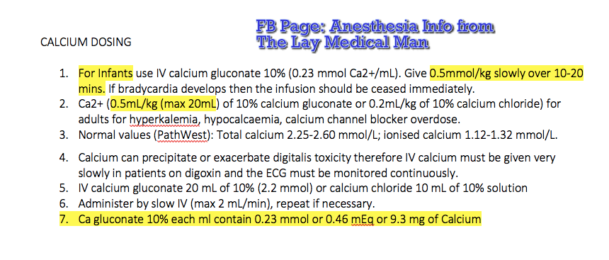
Calcium Gluconate/ Calcium chloride dosing

Screen Shot 2019-08-11 at 3.37.51 pm

VIVA SCENE: GASTRIC ULCER BLEED AND OTHER QUESTIONS

STRESS ULCER RISK FACTORS

Stress ulceration in intensive care patients is relatively common (approaching 90% by day 3 with no prophylaxis), although the incidence of clinically important GI bleeding is less than 2%. There are six major risk factors: 1.Respiratory failure requiring ventilation for >48 hours 2.Coagulopathy 3.Sepsis 4.Hypotension 5.Hepatic failure 6.Renal failure

STRESS ULCER PATHOPHYSIOLOGY

  • Impaired mucosal blood flow 2.Mucosal ischemia 3.Reduced mucus production 4.Reduced mucosal Prostaglandin production 5.increased gastrin production 6.acid-base abnormalities 7.reflux of bile.

TYPES OF ULCERS

Curling’s ulcers associated with extensive burns

Cushing’s ulcers associated with intracranial pathology and gastric acid hypersecretion

STRESS ULCER MANAGEMENT

  • Optimal oxygen delivery to gastric mucosa, avoid hypotension
  • Enteral feeding
  • Proton pump inhibitors, H2 antagonists: by increasing the pH of gastric contents, there is an increased risk of bacterial colonisation and subsequent nosocomial pneumonia.
  • Sucralfate: Aluminium salt of sulphated sucrose and is given in a dose of 1g by NG tube, 6-hourly. It forms a paste at low pH, which preferentially binds to areas of peptic ulceration, thereby providing a physical barrier to the effects of acid. Some investigators have demonstrated a reduction in the rate of nosocomial pneumonias in patients treated with sulcralfate.

MANAGEMENT OF GI BLEED

ECG, NIBP and SpO2 monitoring

Get two wide bore IV access and start RL or NS upto 2 Ls

Support airway: evaluate for the need for airway protection; high volume effective suction must, reduce induction agents dose in hypovolemia

Invasive arterial and central venous pressure monitoring may be necessary in massive bleeds, intubated patients and those with co-morbidities

Insert a Foley for measurement of I/O.

Look for evidence of shock due to severe bleeding: tachycardia,hypotension, ongoing bleed, no response to 2L of crystalloids, HR>90, SBP<100

Rule out hemoptysis and epistaxis

H/O Peptic ulcer disease, NSAID use, alcholism, cirrhosis, coagulation disorders

Order CBC, PT, PTT, BUN, Creatinine, Glucose, Na,K, LFT Cross match 4–6 units of blood as needed

Upright lateral CXR or lateral decubitus abdominal X-rays

Blood should be given promptly if there is persistent haemodynamic instability despite
2 L of crystalloid or colloid, if the initial haemoglobin level is <7 mg/dL, if there is a significant risk of re-bleeding and in those patients with co-morbidities making them unable to tolerate periods of anaemia

Correct coagulopathy

Arrange for endoscopy: both diagnostic / therapeutic

Consider use of vasopressin or octreotide IV, i.v. PPIs.

NSAID INDUCED GASTRIC ULCERS

NSAIDs reduce circulating prostaglandins that are essential in maintaining gastric mucosal integrity. The more an NSAID blocks COX 1, the greater is its tendency to cause peptic ulceration and promote bleeding. Selective COX 2 inhibitors cause less bleeding and fewer ulcers than other NSAIDs

NSAID INDUCED RENAL FAILURE

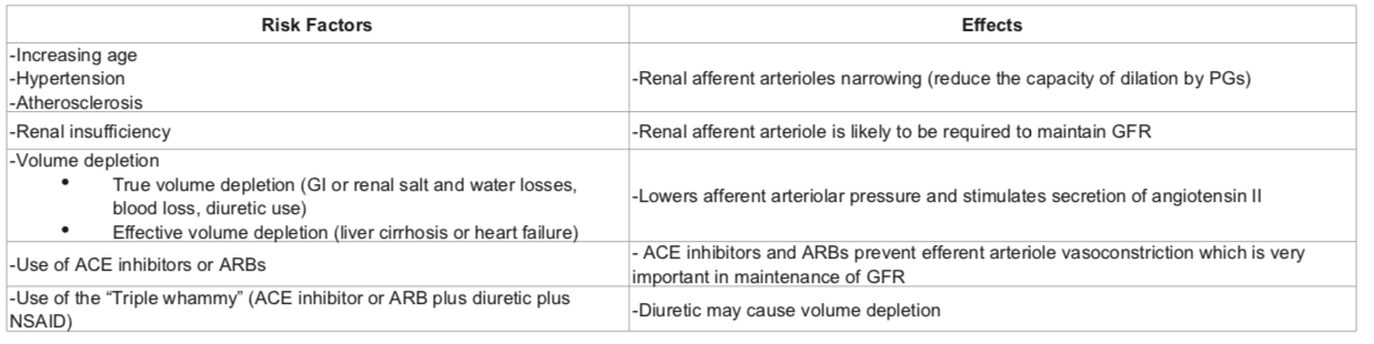
NSAIDs reduce afferent arteriolar blood flow by antagonizing vasodilatory prostaglandins in patients with in patients with risk factors. In this situation, glomerular filtration rate (GFR) drops leading to AKI.

RISK FACTORS: pre-existing renal dysfunction, diabetics, elderly patients, dehydration, decompensated cirrhosis, CHF and patients on ARBs/ACE inhibitors. The mechanism involves reduced levels of prostacyclin, which are required to maintain renal perfusion.

NSAIDs can result in Acute kidney injury (AKI) resulting in the abrupt loss of kidney function, leading to the retention of waste products, electrolyte disturbances, and volume status changes

NSAIDs can also result in Acute interstitial nephritis (AIN) is a renal lesion characterized by a rapid deterioration in kidney function with inflammation and edema of the renal interstitium
Screen Shot 2019-08-10 at 5.10.48 pm.png

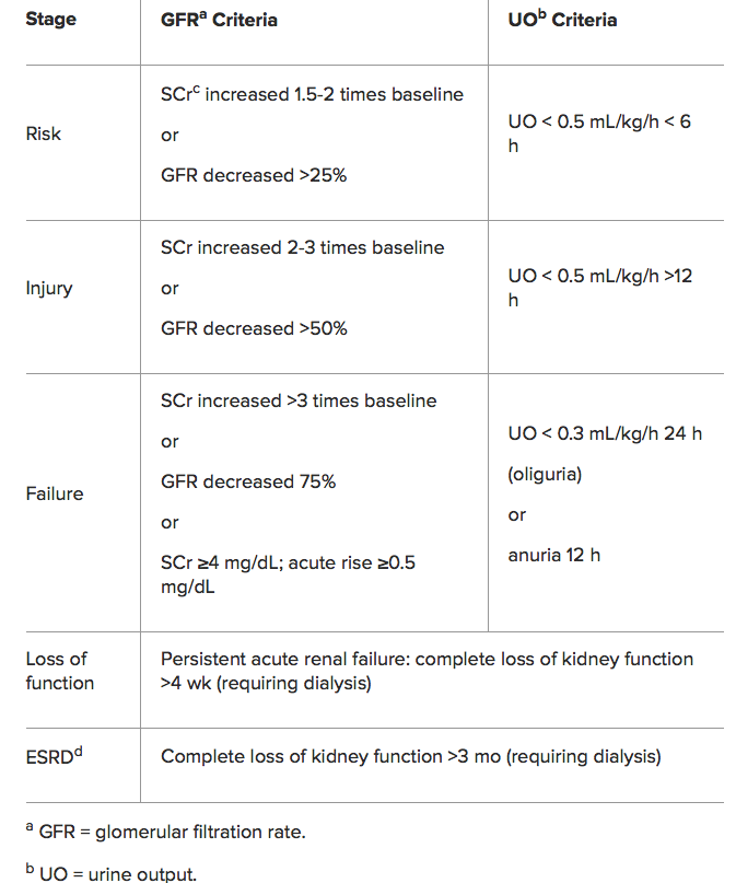
RIFLE Classification for AKI

Screen Shot 2019-08-10 at 5.23.21 pm

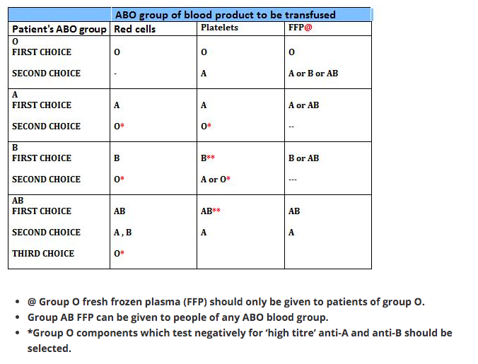
THIS PATIENT REQUIRES BLOOD TRANSFUSION: ABO INCOMPATIBILITY; WHAT’S IT?

Blood groups result from the different antigens expressed on RBCs. ABO and Rh systems are the most important out of the 29 groups. Early in life persons develop antibodies in plasma against non-self antigens.

ANTIGEN IN RBC

ANTIBODY IN PLASMA

Group A has A antigen

Group A has antibody to group B

Group B has B antigen

Group B has antibodies to group A

Group O has no antigen

Group O has antibody to group A and group B

Group AB has A and B antigens

Group AB individuals do not have antibody to group A or B.

So the blood group AB are ideal recipients, as their plasma doesn’t contain any antibody that can agglutinate the donor blood. In the same way blood group O are ideal donors, as once the antibody containing plasma is removed, it contains no antigens to agglutinate with the antibodies in donor’s blood.

RBC

1 Group O individuals can receive blood from group O donors only ( as the antibodies against A or B in their plasma will react with any A or B antigens which enter the circulation)

2 Group A individuals can receive blood from group A and O donors

3 Group B individuals can receive blood from group B and O donors

4 Group AB individuals can receive blood from AB donors, and also from group A, B and O donors ( as their plasma don’t have any antibodies against any antigens)

PLASMA

In plasma transfusion, group AB plasma can be given to a patient of any ABO group because it contains neither anti-A nor anti-B antibody. Group A plasma (with anti-B) can be given to group O and A patients. Group B plasma to group O and B patients only. Group O plasma (anti-A + anti-B) can be given to group O patients only.

PLATELETS

The Platelet Concentrates transfused must be ABO-identical, or at least ABO-compatible, in order to give a good yield (In an emergency, ABO non-identical units can be used, although the improvement seen in platelet count post-transfusion may be less.) Group O PC can be used for patients with blood groups A, B, and AB ONLY IF, they are resuspended in additive/preservative solutions, or if negative for high titre anti-A/A,B. Rh-negative patients, in particular women of childbearing age, should receive, if possible, RhD-negative PC

Rh BLOOD GROUP

Is the second most important group system. Out of the existing C,D and E antigens, D is the most antigenic one. Anti D antibodies are not normally found in the blood of Rh negative individuals; instead they develop it only when itcomes into contact with Rh positive blood during child birth or inappropriate transfusion. In case of subsequent transfusins or pregnancies with Rh positive blood- this can cause rapid destruction of RhD positive red cells (Hemolytic disease of the newborn in subsequent pregnancies; to prevent this sensitization we should give Rhesus imunoglobulin= Anti-D prophylaxis- to the Rh negative mother who gave birth to an Rh positive baby). FFP does not need to be Rh-compatible. Anti-D prophylaxis is not necessary in Rh D-negative recipients of Rh D-positive FFP.
Screen Shot 2019-08-11 at 12.00.04 am

Bradyarrhythmias

Bradycardia is a resting HR < 60 bpm and may be physiological (e.g. in athletes or during sleep), due to intrinsic conduction disease (e.g. heart block), or due to external influences (e.g. cholinergic or adrenergic-blocking medications). With an intrinsic conduction disease, the problem can occur at any point in the SA-AV-His-Purkinje pathway.

PR INTERVAL

The normal PR interval is 120–210 milliseconds (some books say 200 ms!). PR interval is measured from the start of the P wave to the start of the ventricular complex, whether that is a Q or an R wave. At a standard paper speed of 25 mm/s, each small square is equivalent to 40 ms. The normal PR interval is therefore 3–5 (ish) small squares

CAUSES

–> An SA node disease may result in inappropriate sinus bradycardia and pauses either due to failure of the SA node to depolarise or failure of the SA node to capture the surrounding atrial myocardium (sino-atrial exit block).

–> Disease at the level of the AV node will result in alteration in the PR interval or P-QRS relationship. This is often referred to as heart block (HB), of which there are three common types:

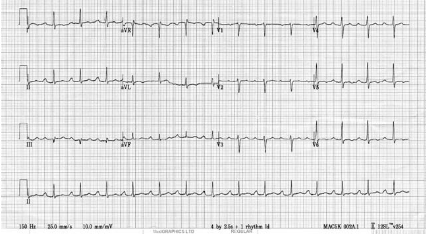
1st degree: Each P-wave is followed by a QRS complex, but the PR interval is prolonged (> 200 ms).

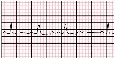
2nd degree: Not all P waves are followed by a QRS complex.

Mobitz Type I: In Mobitz type I (Wenckebach) there is a progressive prolongation of the PR interval (AV conduction) until eventually an atrial impulse is completely blocked. When an atrial impulse is completely blocked there will be a P wave without a QRS complex. This pattern is often referred to as a “dropped beat.”. Here each depolarization results in the prolongation of the refractory period of the atrioventricular node (AVN); hence itis progressive. Eventually, an impulse comes when the AV node is in its absolute refractory period and will not be conducted. This will manifest on the ECG as a P wave that is not followed by a QRS complex. This non-conducted impulse allows time for the AV node to reset, and the cycle continues. When seen in young, fit people with high vagal tone, often nocturnally, it may be benign. However, in the elderly it can be a sign of significant AV node disease and there may be a risk of inducing higher degrees of AV block, particularly with AV nodal blocking agents (e.g. beta-blockers, verapamil, diltiazem, amiodarone).

Mobitz Type II: In Mobitz type II there is a constant PR interval across the rhythm strip both before and after the non-conducted atrial beat. Each P wave is associated with a QRS complex until there is one atrial conduction or P wave that is not followed by a QRS.

3rd degree : There is complete dissociation of P-waves and QRS complexes due to complete block at the AV node (complete HB). The QRS rate is slower than the P-wave rate as the ventricular complexes are generated by an escape rhythm within the ventricular conduction system, which has a lower intrinsic automaticity rate compared to atrial tissue. If the escape rhythm originates high within the His-Purkinje system, the escape rhythm may consist of narrow QRS complexes (since onward ventricular depolarisation occurs via the normal conduction pathway) and only slightly reduced HR (40–50 bpm). The lower the escape rhythm in the His-Purkinje system, the broader and
slower the QRS complexes and the higher the risk of progressing to asystole

MANAGEMENT

–> For bradycardia, in a symptomatic patient give atropine 500 µg IV, and if the patient remains symptomatic, repeat this dose every 3–5 minutes up to a maximum of 3 g. If there has been a calcium antagonist or beta-blocker overdose consider using glucagon. If the patient is at risk of developing asystole, transvenous pacing may be required. Risk factors for a bradycardia deteriorating to asystole include a recent episode of asystole, Mobitz type II AV block, third-degree (complete) heart block or ventricular standstill of more than three seconds.

–> Complete heart block with narrow complexes may not require immediate pacing, as AV junctional ectopic pacemakers can provide a reasonable and stable cardiac output.

–> Transvenous pacing is the definitive treatment, but if the expertise is not readily available, patients may be managed temporarily with either transcutaneous pacing or an epinephrine infusion in the range of 2–10 µg per minute.

–> If there is no pacing equipment immediately available, fist pacing may be attempted. Apply serial rhythmic blows with a closed fist over the left sternal edge to pace the heart at physiological rate of 50–70 times a minute

INDICATIONS FOR TEMPORARY PACING IN THE PERIOPERATIVE PERIOD

  • Complete heart block
  • Second degree heart block
  • First degree heart block with left bundle branch block
  • Trifascicular block

OTHER INDICATIONS FOR TEMPORARY PACEMAKER

  • Bradycardia unresponsive to atropine
  • Inferior wall MI which can impair perfusion to AV node
  • Anterior wall MI affecting bundle branches in the IVS
  • Overdrive pacing for AV reentry tachycardia and VT
  • Post aortic or VSD or tricuspid surgery
  • Asystole with p wave activity

FIRST DEGREE HEART BLOCKScreen Shot 2019-04-23 at 8.37.53 PM.png

SECOND DEGREE HEART BLOCK

Screen Shot 2019-04-23 at 8.38.46 PM

COMPLETE HEART BLOCK

Screen Shot 2019-04-23 at 8.38.13 PM

Broad Complex Tachycardias

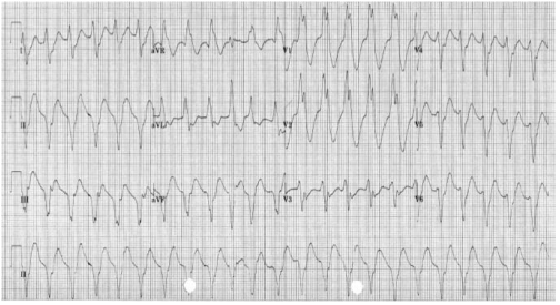
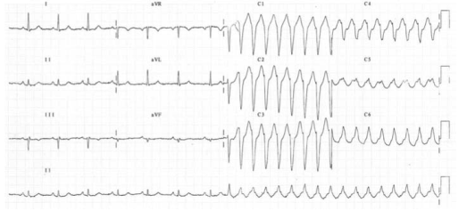
A broad complex tachycardia has a QRS complex greater than 0.12 seconds. They are usually ventricular in origin, but can also be supraventricular with aberrant conduction. Other possible causes for broad complex tachycardias include atrial fibrillation with ventricular pre-excitation, i.e. patients with Wolff–Parkinson–White (WPW) syndrome, or torsades de pointes (polymorphic VT).

Broad complex tachycardia is therefore due to SVT with aberrancy or Ventricular Tachycardia (VT), and differentiating between the two can be challenging. However, there are a few pathognomonic ECG features that diagnose VT

Screen Shot 2019-04-22 at 9.35.57 PM

1.Atrio-ventricular (AV) dissociation. There is a higher ventricular rate than atrial rate (more QRS complexes than P-waves). This can only occur if the ventricular rate is autonomous and no longer under control of the SA node.

2.Capture beats: There is an isolated narrow complex amongst a train of broad complexes. This represents a normally conducted P-wave via the AV node and an intact His-Purkinje system indicating there is no underlying bundle branch block. Therefore, the train of broad complexes are ventricular in origin (i.e. VT).

3.Fusion beats: A normally conducted P-wave may fuse with a simultaneous ventricular beat causing a complex halfway between the appearance of a normal QRS and a broad complex.

4.VT is more likely in patients with a prior history of MI.

5.VT complexes are usually very broad (> 160 ms) due to a very abnormal path taken by the depolarisation wave from the VT focus.

6.The time from R-wave onset to the nadir of the S-wave is prolonged (> 100 ms) in VT, again representing an abnormal activation path through the ventricle.

7.Extreme left axis deviation and positive aVR are more common in VT, as the ventricles are depolarised in the opposite direction to normal conduction.

8.Failure to respond to iv adenosine

9.The absence of typical RBBB or LBBB patterns suggests VT. For example, an RSR pattern in V1 with a taller first R-wave suggests VT (in RBBB the first R-wave is caused by septal depolarisation and is therefore smaller than the second R-wave, which is caused by depolarisation of the RV).

SVT with aberrancy is more likely if previous ECGs demonstrate an accessory pathway or a bundle branch block with identical morphology to the broad complex tachycardia. When in doubt, treat as VT

TORSADES DE POINTES

Is a specific variant of ventricular tachycardia (VT). It has a classic undulating pattern with variation in the size of QRS complex. It is caused by a prolonged QT interval and can precipitate VF and sudden death

QT PROLONGATION: CAUSES

Tricyclic antidepressants, flecainide and quinidine; Hypocalcemia; Acute myocarditis

VENTRICULAR TACHYCARDIA(VT) AND VENTRICULAR FIBRILLATION (VF)

VT is a broad complex tachycardia, defined as a run of at least three consecutive ventricular ectopic beats, at a rate of >120 bpm. Can arise from a single or multiple foci or from a reentry circuit. There may be capture or fusion beats, where a normally conducted beat will join an ectopic beat travelling in the opposite direction

CAUSES: Acute MI, degeneration of other arrhythmias, electrolyte abnormalities etc

VF describes an ECG which is random and chaotic with no identifiable QRS complexes that is incompatible with life and need immediate provision of ACLS with prompt delivery of DC shock. Others: Amiodarone, Lidocaine, beta blockers, Implantable cardioverter defibrillators


MANAGEMENT

For VT treat with amiodarone 300 mg IV followed by 900 mg over 24 hours. If the arrhythmia is known to be supraventricular, treat as a narrow complex tachycardia.

An irregular broad complex tachycardia is most likely to be atrial fibrillation with bundle branch block, and should be treated as narrow complex atrial fibrillation

In a stable patient who is known to have WPW, the use of amiodarone is probably safe. Adenosine, digoxin, verapamil and diltiazem must be avoided, as these drugs block the AV node and will cause a relative increase in pre-excitation

Torsades de pointes is treated by stopping all drugs known to prolong the QT interval and correcting electrolyte abnormalities. Magnesium sulphate (2 g IV over 10 minutes) should also be given. Such patients may require ventricular pacing. If the patient’s condition deteriorates proceed to synchronised electrical cardioversion or, if the patient is pulseless, commence the ALS algorithm

Ventricular bigeminy
Ventricular bigeminy is associated with endotracheal intubation (a sympathoadrenal response). Given time the bigeminy will disappear, but if it does not intravenous
lidocaine (50–100 mg) may be helpful

Narrow Complex Tachycardias

Narrow complex tachyarrhythmias have a QRS duration <0.12 seconds. They arise above the bundle of His.

NARROW COMPLEX TACHYCARDIA

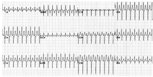
As narrow complex tachycardias involve ventricular activation through the normal His-Purkinje system, they must originate within the atria and are therefore often referred to as supraventricular tachycardia (SVT). There are five common types of SVT. They are: Atrial tachycardia, Atrial fibrillation, Atrial flutter, Atrioventricular nodal
re-entry tachycardia, Atrioventricular re-entry tachycardia. When faced with an ECG of narrow complex tachycardia, (i) we should examine the P-wave and (ii) check the QRS regularity

SINUS ARRHYTHMIA/TACHYCARDIA/BRADYCARDIA (from SA Node)

ATRIAL FIBRILLATION 

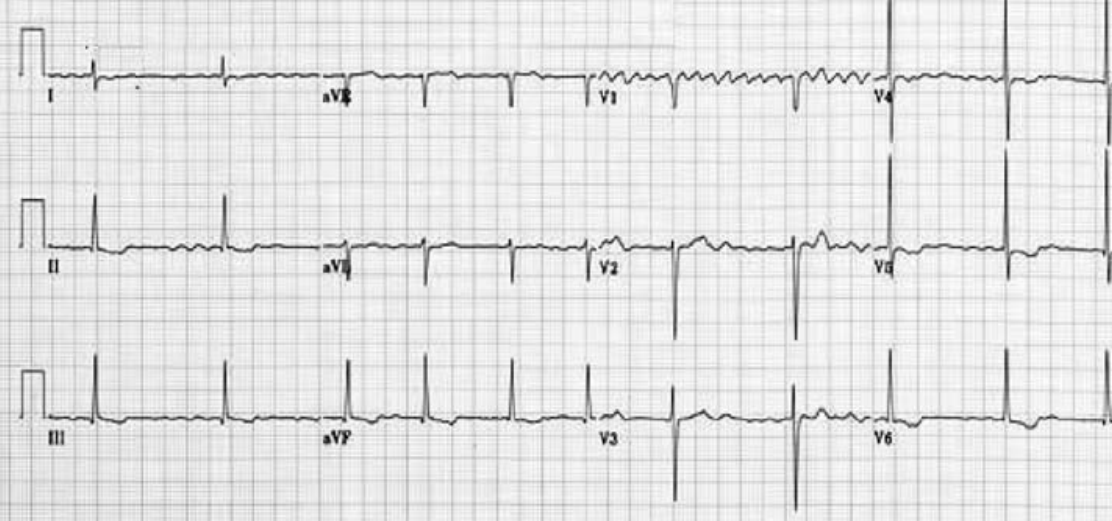
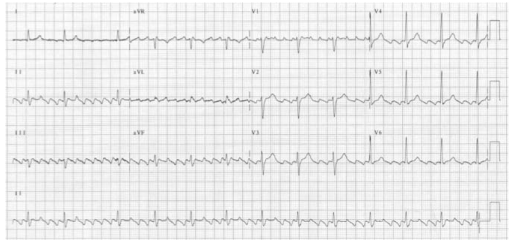
There is completely disorganised atrial activity, with P-waves replaced by an irregular baseline due to fibrillation waves, and QRS complexes occur in an irregularly irregular fashion (Please the post on AF)

Screen Shot 2019-04-18 at 9.23.35 PM

ATRIAL FLUTTER 

There is a self-perpetuating wave of atrial depolarisation usually circulating within the right atrium, causing regular, saw-toothed flutter waves at 300 bpm and QRS complexes every second, third, or fourth flutter wave. We can see classical sawtooth flutter waves.Drug control of the ventricular rate is not often successful.

Screen Shot 2019-04-22 at 8.06.10 AM

ATRIAL TACHYCARDIA 

There is an abnormal atrial focus driving the ventricular rate. This rhythm can be difficult to distinguish from sinus tachycardia, but P-wave morphology and axis is usually abnormal. If the atrial focus is close to the AV node, a junctional tachycardia may occur and P-waves may be absent.

In case of Atrial tachycardia with AV block after halting glycoside therapy (and ensuring normokalaemia), lidocaine 1 mg kg−1 IV is the drug
of choice. Alternatively DC cardioversion or atrial
pacing may be effective.

ATRIO VENTRICULAR NODAL REENTRY TACHYCARDIA (AVNRT)

This is the commonest type of paroxysmal supraventricular tachycardia (PSVT). It is often seen in people without any heart disease, and is usually benign. There is a rapid reentry circuit within the AV node resulting in simultaneous atrial and ventricular depolarisation. The P-wave is usually buried within the QRS or ST-segment. There will be fast regular narrow complex tachycardia, and P-waves can be seen buried in the terminal portion of the QRS complex which may easily be mistaken for a second, small R-wave. The very close proximity of the QRS and P-waves implies near simultaneous depolarisation of atria and ventricles.

Screen Shot 2019-04-22 at 8.23.38 AM

ATRIO VENTRICULAR REENTRY TACHYCARDIA (AVRT)

This occurs in patients with WPW, and is usually benign unless there is coexisting structural heart disease. There is an accessory pathway bridging the atria and ventricles allowing antegrade conduction down the AV node (causing a narrow QRS) and retrograde conduction back to the atria via the accessory pathway. Since the depolarisation wave takes time to complete this circuit, the P-wave occurs after the QRS complex and is often buried within the T-wave. AVRT can occur with antegrade conduction to the ventricles via the accessory pathway, but this will result in ventricular depolarisation via an abnormal route and consequently a broad QRS. In sinus rhythm, antegrade conduction via the accessory pathway produces a short PR interval (as the normal delay in the AV node is avoided) and the abnormal activation of the ventricles produces a slurred upstroke
in the QRS called a delta wave. The QRS complex is said to be pre-excited and can be associated with repolarisation abnormalities. There are seven sinus beats followed by a ventricular ectopic beat that conducts to the atria retrogradely through the atrioventricular node and then returns to the ventricles via the accessory pathway. This cycle repeats and triggers a broad complex tachycardia. (Please see post on ‘WPW Syndrome’ also).

Screen Shot 2019-04-22 at 8.44.52 AM

An unstable patient presenting with a regular narrow complex tachycardia should be treated with electrical cardioversion. If this is not immediately available, adenosine should be given as a first-line treatment. A stable patient presenting with a regular narrow complex tachycardia should initially be treated by vagal
manoeuvres such as carotid sinus massage or the Valsalva manoeuvre, as these will terminate up to a quarter of episodes of PSVT. Carotid sinus massage should be avoided
in the elderly, especially if a carotid bruit is present, as it may dislodge an atheromatous plaque and cause a stroke

Management

A stable patient presenting with a regular narrow complex tachycardia should initially be treated by vagal manoeuvres such as carotid sinus massage or the Valsalva manoeuvre, as these will terminate most episodes of PSVT. Carotid sinus massage should be avoided in the elderly, especially if a carotid bruit is present, as it may dislodge an atheromatous plaque and cause a stroke. If the tachycardia persists and is not atrial flutter, 6 mg of adenosine should be given as an IV bolus, followed by a 12 mg bolus if no response. A further 12 mg bolus of adenosine may be given if the tachycardia persists. Vagal manoeuvres or adenosine will terminate almost all AVNRTs or AVRTs within seconds, and therefore failure to convert suggests an atrial tachycardia such as atrial flutter. If adenosine is contraindicated, or fails to terminate a narrow complex tachycardia, without first demonstrating it as atrial flutter, give a calcium-channel blocker, e.g. verapamil 2.5–5 mg IV over two minutes. Atrial flutter should be treated by rate control with a beta-blocker.

An irregular narrow complex tachycardia is most likely to be atrial fibrillation (AF) with an uncontrolled ventricular response, but may also be atrial flutter with variable block. If the patient is unstable, synchronised electrical cardioversion should be used to treat the arrhythmia